


 10 / 17

Page 37
December 9-10, 2019 | Barcelona, Spain
Volume 14
ARTHRITIS AND RHEUMATOLOGY
ANATOMY AND PHYSIOLOGY
13
th
International Conference on
3
rd
International Conference on
&
Journal of Orthopaedics Trauma Surgery
and Related Research
Rheumatology Congress 2019 & Anatomy and Physiology 2019
December 09-10, 2019
J Orthop Trauma Surg Rel Res, ISSN: 1897-2276
High number of negative radiographs for suspected tibial shaft fracture adds expense
and increases patient throughput time in the emergency department
Safa Fassihi
George Washington University Hospital, USA
Purpose
: The diagnosis of tibial shaft fractures (OTA 42A-C) is commonly made by emergency department (ED) providers prior
to orthopedic consultation. Due to the subcutaneous anatomy of the tibia, a comprehensive history and physical examination
are often sufficient for fracture diagnosis, with radiographs serving as a secondary aid. A high rate of negative X-Rays increases
cost and inefficiency in the ED. This study aims to define the rate at which tibial radiographs are negative for suspected fracture.
Methods
: At a Level I trauma center, a prospective database was retrospectively evaluated for ED radiographs taken from 2014
to 2016. Only radiographs obtained for suspected fracture of the tibial diaphysis were included. From this, the rate of negative
diagnostic studies and the associated costs, ED throughput time, resource utilization, and radiation exposure were calculated.
Results
: During the study period, 734 tibia radiographs were performed for diagnosis of tibial shaft fracture without suspected
adjacent articular pathology. Of these, 565 (76.9%) were negative for tibial shaft fracture. Patient charges were increased by these
radiographs through both higher radiology charges ($598 per tibia radiographic
series) and higher professional charges. The mean time to obtain a tibia X-ray series
in the ED was 57 minutes (SD: 47 minutes; Median: 47 minutes). The radiation
exposure from a tibia radiographic series was 15 millirems.
Conclusion
: At this institution, a large proportion of the radiographs obtained for
suspected tibial shaft fracture are negative. The resources and time spent acquiring
these radiographs places higher demands on physicians and staff while increasing
charges and radiation exposure to patients. In addition, these negative radiographs
add throughput time in the ED, thereby potentially contributing to ED overcrowding.
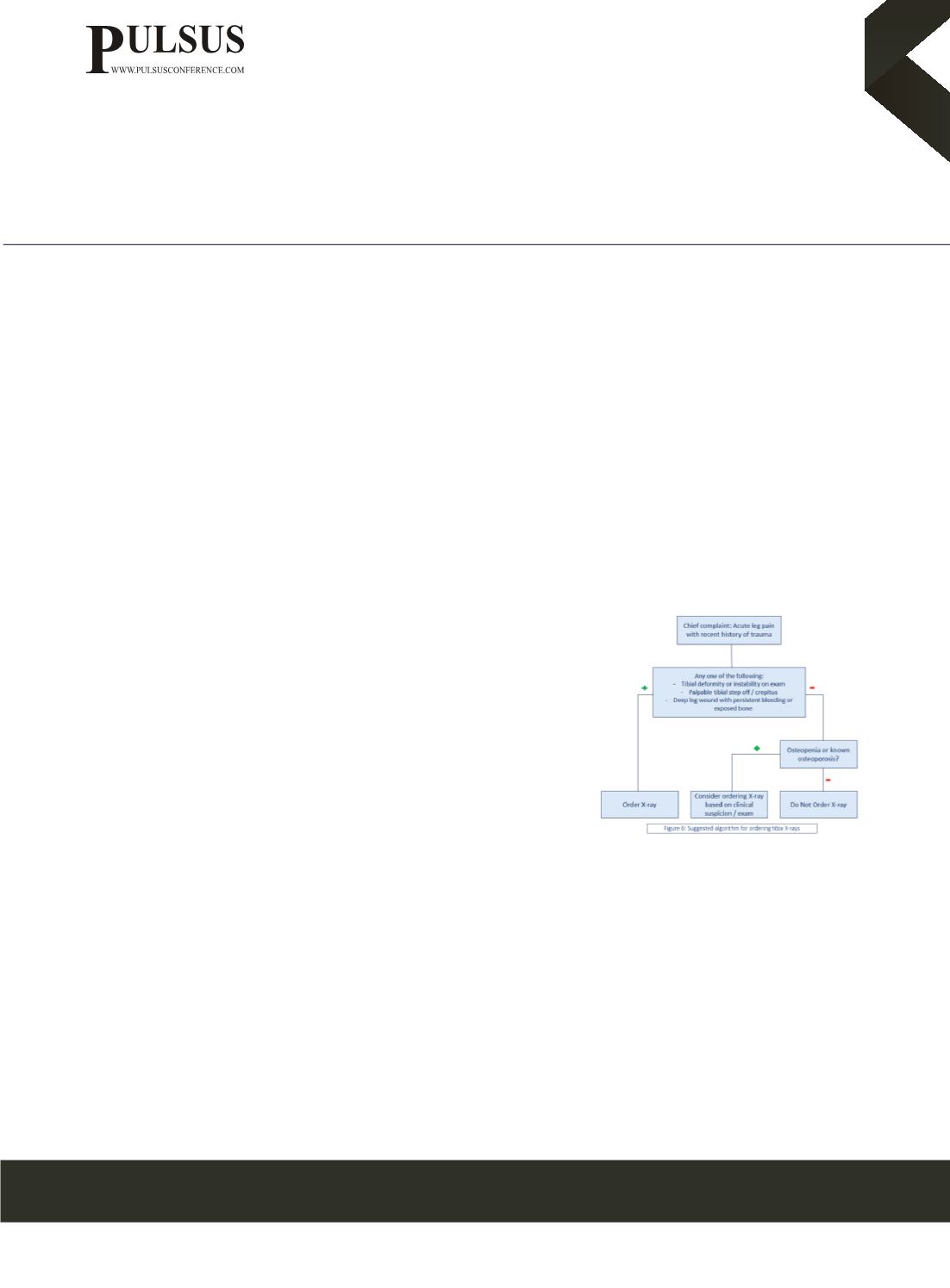
The authors propose a systematic approach to maximize the diagnostic efficiency of
tibia radiographs and subsequently improve resource allocation in the ED.
Biography
Safa Fassihi is a US-based physician pursuing a career in orthopedic total joint arthroplasty. His research focuses not only on total joint
arthroplasty but also on improving the procedural and financial efficiency of the healthcare system. This specific analysis was based
upon his experience at a level 1 trauma center, in which he recognized that there was significant emergency department overcrowding
on a regular basis, so he sought to minimize any factors that may be contributing to this problem. He noticed that tibia X-rays were
frequently ordered for suspected tibial shaft fracture and were negative most of the time. He collaborated with a US board-certified
orthopedic traumatologist to identify the extent of this problem and offer a potential solution.
scf5071@gmail.com