Orthoplastic consideration during distal tibia deformity and limb lengthening utilizing truelok hexapod circular external fixator
2 Director, Foot and Ankle Specialist of Ohio, Reconstructive Surgery and Deformity Correction Fellowship Program, Ohio, USA
3 Department of Orthopedics, United States Naval Hospital Yokosuka, Japan
4 Chicago Foot and Ankle Deformity Correction Fellow, USA
Received: 21-Nov-2017 Accepted Date: Jan 16, 2018 ; Published: 19-Jan-2018
This open-access article is distributed under the terms of the Creative Commons Attribution Non-Commercial License (CC BY-NC) (http://creativecommons.org/licenses/by-nc/4.0/), which permits reuse, distribution and reproduction of the article, provided that the original work is properly cited and the reuse is restricted to noncommercial purposes. For commercial reuse, contact reprints@pulsus.com
Abstract
Background: This case study examines the utilization of Hexapod Truelok circular external fixation as well as the osteotomy site with respect to avoid further damage to the cutaneous perforators on patients that had a poor soft tissue envelope with performing angular lower limb deformity correction within the 55-year-old to 74-year-old age group with underlying comorbidities.
Methods: A prospective study was done on 17 patients with angular tibial/fibular deformities. 9 of them were males and 8 of them were females. Fixation of all cases was performed with utilization of Hexapod Truelok external ring fixation with injection of bone marrow 9X concentrate drawn from the tibia to the osteotomy region during the initial surgical intervention. An additional peripheral blood along with bone marrow concentrate with fluoroscopic percutaneous guided injection was placed into the osteotomy/lengthening site during the post-operative period when the footplate was removed.
Results: Circular Hexapod Truelok external fixation was applied for an average of 17 weeks post-operatively with no non-union, mal-union or infection.
Conclusion: Gradual correction for distal tibial and fibular osteotomy with combined orthoplastic approach allows for improved outcomes to be obtained. Hexapod Truelok application with external ring fixation is a useful alternative with correction of a multiplane deformity simultaneously in an easily applied prescription principle format.
Keywords
Distal tibia and fibular osteotomies, Tibial and foot deformities, Truelok external ring fixator, Hexapod external ring fixator, Angular limb deformity, Osteoplastic Orthoplastics
Introduction
With the variety of fixation devices available to correct multiplane deformities; Hexapod Truelok circular external fixation is a relatively sound method for fixation with an adaptable prescription base adjustment approach. Hexapod Truelok external fixation with gradual correction has been proven effective in correcting multiplanar tibial deformities simultaneously [1-5].
Hexapod Truelok fixation allows a unique prescription to be applied to the correct corresponding strut(s) to adjust the surgical limb to its corrective length and/or apply an angular correction with 1 mm increment fine-tuning knobs [6]. This case study is to evaluate the ease and effectiveness in treatment of angular limb deformities with having optimal results.
Patients and Methods
A case study was done on 17 patients with angular limb deformities of the tibia with underlying comorbidities during 2015 to 2017. External fixation was in place for an average of 17 weeks post operatively with 0.5mm/ day with incremental adjustment twice per day. 9 were males and 8 were females.
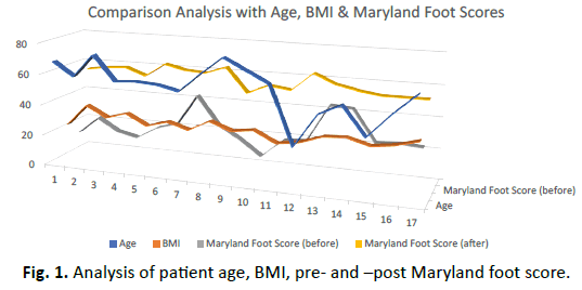
Age, laterality, associated injury(s), days of correction, type of procedure, comorbidities, parameters of deformity were documented. All patients underwent surgical intervention and lower limb lengthening with application of external ring fixators with usage of Hexapod Truelok struts (Table 1 and Fig. 1).
| Variables | Values |
|---|---|
| Sex | 9 males, 8 females |
| Age | 55-74 |
| Laterality | 9 RT 8 LT |
| BMI | 23 – 34 |
Table 1. Patient demographics.
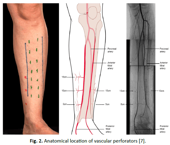
All patients were placed under general anesthesia without the usage of a tourniquet. With the patient within the supine position, the lower limb is prepped and draped in the aseptic manner. Anatomical landmarks are placed to the tibial tuberosity, tibial crest, ankle joint, distal fibula with side mark midline of the fibula, lateral process of the talus, distal 2nd interspace (dorsally and plantarly) and plantar bisection of the calcaneus. Prior to performing the frame application & tibial osteotomy, identification of the 5-10-15 cm perforators was performed with a handheld doppler to avoid vascular impairment overlying the medial, anterior and lateral compartment of the lower leg [7] (Fig. 2). Attention was brought to have pristine alignment of the circular external fixator to reduce software data entry. The constructed ring fixator is assembled for a total of 4 rings with 2 full circular rings, 5/8 ring and foot support with the opening facing anteriorly (described from proximal to distal). The hexapod support is constituted to the 2nd and 3rd proximal rings which allows the surgeon to create a virtual hinge to simultaneously correct complex multi-planar deformities without the need to alter the frame construct [8].
Fig 2. Anatomical location of vascular perforators [7].
The constructed circular fixator is placed with the center of the rings parallel to the tibial crest and the proximal ring being 3 cm below the tibial crest, 5/8 circular ring 1 cm proximal to the ankle joint and the foot plate 1 cm distal to the distal lateral malleolus. When aligning the lower leg within the external fixator, it’s important to also have the side mark midline to the bisection of the fibula and the leg fully extended. A single half pin is placed on the first and second proximal ring anteriorly, medial to the tibial crest trajecting 10° from their respective ring towards one another. Such placement of the half pins avoids injury to the perforators.
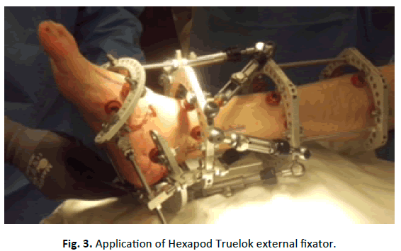
Placement of the half pins stabilizes the alignment of the proximal two rings with confirmation of anatomic alignment to the external fixator. Once confirmed, two trans-osseous fixation wires at 60° are thrown along the 1st and 2nd proximal rings. Two olive wires are then placed 60° to one another within the proximal 3rd ring with retrospective alignment to the bisection line of the fibula. A foot support plate is attached with two olive wires at 60° to one another into the calcaneus and two smooth wire angled at 60° to each other in the midfoot. With placement of the footplate wires, it is imperative to maintain rectus alignment of the heel bisection line and the 2nd interspace markings to be in alignment with the calcaneus bisection and tibial crest. The foot support is a stabilizer to avoid equinus of the ankle post operatively (Fig. 3) [9].
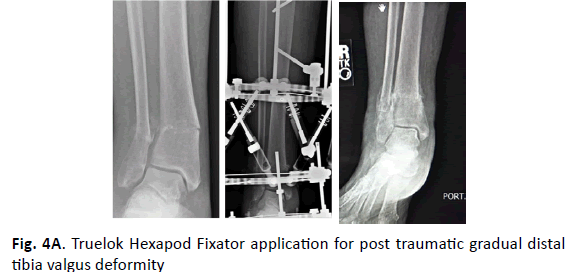
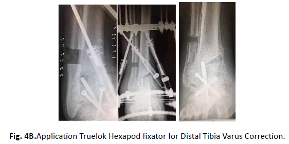
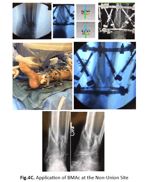
Corticotomy was performed under fluoroscopic guidance utilizing the classic Ilizarov technique minimizing soft tissue exposure and blood loss by a 1cm linear incision intermediate to the 5 and 10 cm perforators. With performing the corticotomy, the osteotome does not penetrate the medulla and periosteum. This gives the surgeon the opportunity to adjust the alignment and/or compress/distract after surgery (Fig. 4A-C). All patients prior are evaluated intraoperatively to preserve the perforators of the anterior tibial and peroneal arteries to avoid damage at the time of corticotomy in order to preserve the soft tissue envelope [2]. The incision placement of approximately 1cm in length is placed parallel to the tibial crest within the medial compartment of the tibia and along the lateral bisection line of the fibula intermediate to the 2nd and 3rd proximal ring construct.
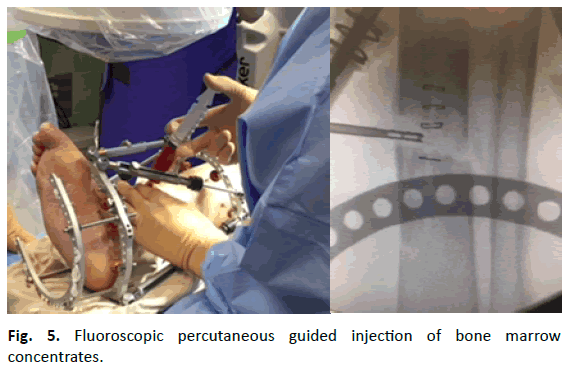
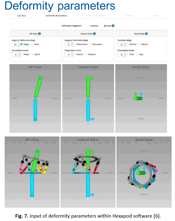
Injection of bone marrow concentrate (9X concentration) with 60 cc drawn from the proximal tibia is placed along the osteotomy sites with fluoroscopic guidance. The patient is placed on partial weightbearing status of 20% immediately post operatively. The patients are given a prescription on their respective strut adjustments to be performed twice per day for distraction and angular correction of 0.5mm/day increments. With usage of software prescription adjustment(s), the hexapod circular external fixator can accurately correct deformities and limb-length discrepancies simultaneously. (10) All patients were monitored with serial radiographs (Fig. 5-7).
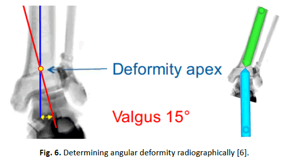
Fig 6. Determining angular deformity radiographically [6].
Fig 7. Input of deformity parameters within Hexapod software [6].
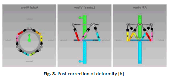
Once the desired correction is achieved, the patient is brought back to the operating room for removal of the foot support along with fluoroscopicpercutaneous injection of 3 cc concentrate to the osteotomy sites using 60 cc of peripheral blood concentrate spun down by using a Magellan Isto device (Fig. 8). The external ring fixator remains in place for a minimum amount of days equal to (2 × days of distraction) + days of latency.
Fig 8. Post correction of deformity [6].
Final removal of the external ring fixator is determined after CT tomography reveals adequate consolidation. After the fixator is removed, the patient is placed within a patellar tibial fracture orthoses for a period of 8 weeks with incremental weightbearing status per rehabilitation instructions. Rehabilitation is ordered to mobilize the knee, ankle, subtalar and midtarsal joints post operatively.
Result and Discussion
17 cases of angular lower limb and distraction were corrected with usage of Hexapod Truelok circular external fixation including 9 males and 8 females. Age was from 55 years to 74 years. There were 9 right, 8 left. Corticotomy Ilizarov osteotomy was chosen over open power saw osteotomy because patients had poor soft tissue envelopes and avoidance to further damage their cutaneous perforators. All patients prior to osteotomies had their perforators of the anterior tibial and peroneal arteries identified to avoid damage at the time of the corticotomy. Gradual correction was chosen over acute correction to prevent damage to the soft tissue envelope avoiding larger incision approach that would damage cutaneous perforators involving the 3 main angiosomes of the lower limb [2,5]. Bone marrow aspirate with mesenchymal stem cells were utilized along the osteotomy site to improve healing with reduction in non-unions [10]. There were 3 patients with (18%) insulin dependent with peripheral neuropathy, 1 (6%) with non-insulin dependent with peripheral neuropathy, 15 (88%) with secondary osteoarthritis of the ankle, 6 (35%) with previous history of distal tibial fractures, 8 (47%) with intrinsic ankle varus, 2 (12%) with Charcot arthropathy, 2 (12%) with congenital clubfoot deformity, 5 (29%)with distal tibial valgus deformity and 1 (6%) with fibular hemimelia. Circular external fixation was applied for an average of 17 weeks postoperatively prior to removal with no non-union, mal-union or infection. Hexapod fixators are well suited for harnessing the healing potential of the strain/tension-stress principle to optimize bone and soft tissue healing [11].
Conclusion
Following of gradual correction for distal tibial and fibular osteotomies with combining orthoplastic approach to avoiding damage to the local perforators, good functional outcomes can be obtained with usage of a hexapod external ring fixator.
REFERENCES
- Rodriguez E.R., Maria M.L.: Combined use of the Ilizarov method, concentrated bone marrow aspirate (cBMA), and platelet-rich plasma (PRP) to expedite healing of bimalleolar fractures. Strategies in Trauma and Limb Reconstruction. Strategies Trauma Limb Reconstr. 2015;10:161-166.
- Taylor G.I., Corlett R.J., Ashton M.W.: The functional angiosome: Clinical implications of the anatomical concept. Plastic and reconstructive surgery. 2017;140:721-733.
- Abuomira I.E., Sala F., Elbatrawy Y., et al.: Distraction osteogenesis for tibial nonunion with bone loss using combined Ilizarov and Taylor spatial frames versus a conventional circular frame. Strategies in Trauma and Limb Reconstruction. 2016;11:153-159.
- Christopher I.: Limb lengthening combined with deformity correction in children with the Taylor spatial frame. Journal of Pediatric Orthopaedics. 2010;B19:529-534.
- Pesenti S., Lobst C.A., Launay F.: Evaluation of the external fixator Truelok Hexapod System for tibial deformity correction in children. 2017;103:761-764.
- Orthofix Distal Tibia Pre-operative Planning Tutorial. TLhex.com. [Online] [Cited: 11 07, 2017.] Rights granted from Texas Scottish Wright Children's Hospital for hexapod software images. http://www.tlhex.com/downloads.php?tk=EPRvOBzPBNjLv0Ip3qWqEjO__GL7N1Xj25x7263mzDekbAGzGAXyRwZaFbNGql6HfF7Bc4c1jc4oabVyCuOZu2GhWLJroT1CTZUb5LM8GNk0h_p3udb21AO17UpMGvfo0sEf6ewVO44gvIGvo3_Dlg2.
- Nanchahal J.: Compartment Syndrome. Standards for the management of open fractures of the lower limb. British Association of Pastic Reconstructive and Aesthetic Surgeons. 2009;13.
- Ferreira N., Birkholtz F.: Radiographic analysis of hexapod external fixators: Fundamental differences between the Taylor Spatial Frame and TrueLok-Hex. Journal of medical engineering & technology. 2015;39:173-176.
- Mikhail S.L.: Stepwise approach to equinus deformity correction with circular external fixation. Musculoskeletalkey.com. [Online] August 2, 2016. https://musculoskeletalkey.com/stepwise-approach-to-equinus-deformity-correction-with-circular-external-fixation/.
- Ferreira N., Marais L.C., Aldous C.: Hexapod external fixator closed distraction in the management of stiff hypertrophic tibial nonunions. The Bone & Joint Journal. 2015;97-B:1417-1422.
- Mahomed N., O'Farrell P., Barnard A.C., et al.: Monofocal distraction treatment of stiff aseptic tibial nonunions with hexapod circular external fixation. Journal of Limb Lengthening & Reconstruction. 2017;3:101-106.



 Journal of Orthopaedics Trauma Surgery and Related Research a publication of Polish Society, is a peer-reviewed online journal with quaterly print on demand compilation of issues published.