Is there a difference in femur length between neglected developmental dysplasia of the hip and contralateral normal hip-femur length a radiographic study
2 Reconstructive Orthopedic Department, King Fahad Medical City, Riyadh, Saudi Arabia
3 College of Medicine, King Saud Bin Abdulaziz University for Health Sciences (KSAUHS), Riyadh, Saudi Arabia
4 Department of Orthopaedic Surgery, College of Medicine, Al-Maarefa University, Riyadh, Saudi Arabia
5 Department of Orthopaedic Surgery, Dallah Hospital, Riyadh, Saudi Arabia
Received: 04-Apr-2022, Manuscript No. jotsrr-22- 59379; Editor assigned: 05-Apr-2022, Pre QC No. jotsrr-22- 59379 (PQ); Accepted Date: Apr 25, 2022 ; Reviewed: 18-Apr-2022 QC No. jotsrr-22- 59379 (Q); Revised: 20-Apr-2022, Manuscript No. jotsrr-22- 59379 (R); Published: 26-Apr-2022, DOI: DOI.10.37532/1897- 2276.2022.17(4).72
This open-access article is distributed under the terms of the Creative Commons Attribution Non-Commercial License (CC BY-NC) (http://creativecommons.org/licenses/by-nc/4.0/), which permits reuse, distribution and reproduction of the article, provided that the original work is properly cited and the reuse is restricted to noncommercial purposes. For commercial reuse, contact reprints@pulsus.com
Abstract
Background: There are several anatomical problems with the hip joint that are associated with Developmental Dysplasia (DDH), such as the femoral head being out of place about the acetabulum. First-born status, female sex, a positive family history, breech presentation, and oligohydramnios are all risk factors for preterm labor and birth. DDH severity has been graded using a variety of classification systems, including the Crowe classification, the Hartofilakidis classification, and the Eftekhar and Kerboul classification. The purpose of this study was to determine whether there is a difference in femur length between patients with neglected developmental dysplasia of the hip (DDH) and the normal femur.
Materials and Methods: This is a case series study of 14 patients with Unilateral DDH who did not have surgery. Between January 2017 and December 2020, data were retrieved and obtained from our hospital’s picture archiving and communication system (PACS). A Pelvis x-ray and a Full-Length Femur x-ray were taken for those patients. As a radiological landmark, a full-length film from the tip of the greater trochanter to the intercondylar space was used in this study. The following were the inclusion criteria: 1. The patient must be an adult who is at least 18 years old. 2. The deformity should only occur on one side (Unilateral DDH). 3. They had never had surgery before. 4. Crowe types III and IV
Result: The mean age of the patients was 34 (SD 12.4) years, with females outnumbering males (71.4% vs 28.6 %). Additionally, the mean length of the affected femur was 41.6 (SD 3.88), and the mean length of the normal femur was 42.2. (SD 4.08). When we compared the baseline characteristics of patients by age group (35 years vs 35 years), we discovered that the BMI of the older age group (35 years) was statistically significantly higher than the younger age group (35 years) (P-value =0.028) Result: The mean age of the patients was 34 (SD 12.4) years, with females outnumbering males (71.4 % vs 28.6 %). Additionally, the mean length of the affected femur was 41.6 (SD 3.88) And the mean length of the normal femur was 42.2. (SD 4.08). When we compared the baseline characteristics of patients by age group (35 years vs 35 years), we discovered that the BMI of the older age group (35 years) was statistically significantly higher than the younger age group (35 years) (P-value=0.028).
Conclusion: As a result of our study, we found an approximately 1cm to 2 cm difference in femur length between patients with unilateral DDH and normal hip, which was correlated with age and body mass index (BMI). Preoperative considerations for unilateral DDH include taking a long film of both femurs to determine their relative length differences. This will assist in determining the amount of subtrochanteric femoral osteotomy to perform.
Keywords
Developmental Dysplasia of the Hip (DDH), Femur Length, Crowe Classification, Body Mass Index (BMI)
INTRODUCTION
Developmental Dysplasia of the Hip (DDH) is a group of anatomical abnormalities of the hip joint in which the femoral head has an abnormal relationship with the acetabulum [1]. The incidence ranges from 1 in 1000 to 34 in 1000. When ultrasonography is utilized in conjunction with a clinical evaluation, a higher incidence is reported [2,3]. Firstborn status, female sex, a positive family history, breech presentation, and oligohydramnios are all risk factors i.e. supplementary to clinical examination.
DDH can be divided into three types in adults:-
Type I: Dysplasia in which the femoral head remains in the real acetabulum
Type II: Low dislocation where the femoral head articulates with a false acetabulum covering a partially real acetabulum
Type III: High dislocation in which the femoral head migrated superior posteriorly afterward is not in real acetabulum contact [4-7]. There are different classification systems, including the Crowe classification, the Hartofilakidis classification, and the Eftekhar and Kerboul classification, have been used to grade the severity of DDH [8]. The Crowe classification is the most frequently used in literature. To classify the value of femoral head displacement, the Crowe classification considers the distance between the femoral head center and the inferior margin of the acetabulum [9]. Radiological criteria for DDH vary in the literature, but parameters are generally accepted as Central-Edge (CE) angles <20° and acetabular angles >47° [10,11]. However, unexpected long femurs have been observed in adults with DDH who were not treated surgically as children. We noticed that a patient who had unilateral DDH crow IV even though we had done osteotomy and shortening of approximately 6 cm had a crow IV. Following the operation, we noticed that the femur in neglected DDH is significantly longer than normal. The purpose of this study was to determine whether there is a difference in femur length between patients with neglected Developmental Dysplasia of the Hip (DDH) and the normal femur.
Methodology
This study was conducted at Prince Sultan Military Medical City (P.S.M.M.C), a tertiary care facility in Riyadh, Saudi Arabia. Our institution’s Institutional Review Board approved this study. Between January 2017 and December 2020, data were retrieved and obtained from our hospital’s Picture Archiving and Communication System (PACS). For those patients, a Pelvis X-ray and a Full-Length Femur X-ray were performed. The following criteria were used to determine inclusion in our study:
1. The patient must be an adult and at least 18 years old and above 2. The deformity should be on one side (unilateral DDH) 3. They had no prior surgery 4. Crowe type III & IV
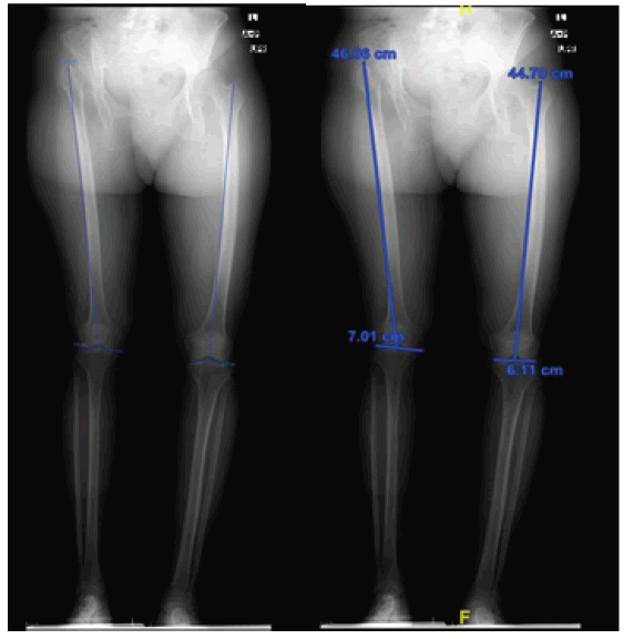
The full-length film from the tip of the greater trochanter to the intercondylar space was used in this study as a radiological landmark (Figure 1).
Statistical Analysis
The data analyses were performed using the statistical package for social sciences, version 26 (SPSS, Armonk, NY: IBM Corp.). Categorical variables were presented using numbers and percentages while continuous variables were presented using mean and standard deviation. Paired t-test was performed to determine the differences in a mean between normal and affected femur length. Furthermore, the normal and affected femur length was compared to the different characteristics of the patients by using an independent sample t-test and Fischer Exact test. Correlation procedures were also conducted to determine the linear relationship between the normal and affected femur length concerning BMI and age. P-value<0.05 was considered statistically significant.
Results
We analyzed 14 patients who were diagnosed with DDH. The mean age of the patients was 34 (SD 12.4) years with females dominating the males (71.4% vs 28.6%). The prevalence of patients with a family history of DDH was 28.6%. Furthermore, class 3 Crowe classification constitutes 64.3% while class 4 constitutes 35.7%. The mean values of weight(kg), height(cm) BMI (kg/m2 ) were 65.5, 155.5 and 28 respectively. In addition, the mean value of the affected femur length was 41.6 (SD 3.88) and the mean value of the normal femur length was 42.2 (SD 4.08). Paired T-test was performed to determine the differences in length between the normal and affected femur. Based on the results, it was found that the length of the affected femur was statistically significantly higher than the normal femur length. (Mean diff.: -0.596; 95% CI: -1.140 to -0.051; p=0.034) The comparison of femur length concerning gender, family history, and Crowe classification was presented. Our investigation revealed that there was no significant difference being observed among gender, family history of DDH, and Crowe classification in both normal and affected femur length (all p>0.05). When conducting correlation procedures between age in years and BMI in regards to normal and affected femur length, it was observed that the correlation between age in years and BMI about normal and affected femur did not reach statistical significance (p>0.05). When comparing the baseline characteristics of the patients in regards to the age group (age<35 years vs ≥ 35 years), we know that the BMI of the older age group (≥ 35 years) was statistically significantly higher than younger age group (<35 years) (p=0.028). Other baseline characteristics of the patients did not significantly influence when compared to age group (p>0.05)
Discussion
When a unilateral dysplastic hip is present, the affected femur is frequently longer than expected. Metcalfe et al. in their study found to have 66% of patients had unilateral DDH, and the femur length was longer than the normal femur with a peak frequency in the 5 mm10 mm compared to the bilateral group [12]. Rai et al. in their study of the discrepancy in the length of the Tibia in unilateral congenital dislocation of the hip [13]. They included 10 patients who had unilateral DDH. They used a reference point from the medial joint line of the knee and the tip of the medial malleolus. The average tibia shortening on the affected side was 1 cm, and it was unrelated to the dislocation severity.
In our study, we found a clinically significant difference in femur length between affected and normal hips. Crowe type III accounts for the vast majority of them (64.3%). Furthermore, there was a clinical correlation between BMI in patients over the age of 35.
We focused on femur length in this study because we typically perform surgery on Crowe type III and IV patients. They all require a sub trochanteric femoral osteotomy, so the difference will give us how much shortening is required to reduce the femur into the native acetabulum. However, in these cases, soft tissue release is required, and the results will influence the decision on pre-operative planning. There were two potential flaws in this study that could have skewed the results. For starters, the chosen sample size was insufficient when compared to the study’s intended audience. Second, only one instrument was used to gather the data for the study. The instruments’ validity and reliability haven’t been thoroughly investigated. Because of the scoring system, there is a high risk of bias affecting the results’ accuracy and reliability.
Conclusion
There is insufficient evidence to support our hypothesis. However, we need to conduct additional studies on bone length. Our study demonstrated a difference in femur length between unilateral DDH and normal femurs of approximately 1 cm-2 cm, which was correlated with the patient’s age and BMI. We recommend that as part of preoperative planning for unilateral DDH, a long film of both femurs be taken to determine the length difference between them. This will assist in determining the amount of sub-trochanteric femoral osteotomy to perform.
References
- Amstutz H.C., Le Duff M.J., Campbell P.A.: Clinical and radiographic results of metal-on-metal hip resurfacing with a minimum ten-year follow-up. J Bone Joint Surg Am. 2010;92:2663-71.[Google Scholar] [Crossref]
- Klein G.R., Levine B.R., Hozack W.J., et al.: Return to athletic activity after total hip arthroplasty. Consensus guidelines based on a survey of the Hip Society and American Association of Hip and Knee Surgeons. J Arthroplasty. 2007;22:171-175. [Google Scholar][Crossref]
- McKee G.K., Watson-Farrar J.: Replacement of arthritic hips by the McKee-Farrar prosthesis. J Bone Joint Surg Br. 1966;48:245-259. [Google Scholar] [Crossref]
- Barrack R.L., Ruh E.L., Berend ME.: Active patients perceive advantages after surface replacement compared to cementless total hip arthroplasty? Clin Orthop Relat Res. 2013;471:3803-3813. [Google Scholar] [Crossref]
- Lavigne M., Masse V., Girard J., et al.: Return to sport after hip resurfacing or total hip arthroplasty: a randomized study. Rev Chir Orthop Reparatrice Appar Mot. 2008;94:361-367. [Google Scholar] [Crossref]
- Carrothers A.D., Gilbert R.E., Jaiswal A., et al.: Birmingham hip resurfacing: the prevalence of failure. J Bone Joint Surg Br. 2010;92:1344-1350. [Google Scholar] [Crossref]
- Su E.P., Su SL.: Surface replacement conversion: results depend upon reason for revision. J Bone Joint. 2013;95:88-91. [Google Scholar] [Crossref]
- Shimmin A.J., Bare J., Back D.L.: Complications associated with hip resurfacing arthroplasty. Orthop Clin North Am. 2005;36:187-193. [Google Scholar] [Crossref]
- Glyn-Jones S., Pandit H., Kwon Y.M., et al.: Risk factors for inflammatory pseudotumour formation following hip resurfacing. J Bone Joint Surg Br. 2009;91:1566-1574. [Google Scholar] [Crossref]
- Jolley M.N., Salvati E.A., Brown G.C.: Early results and complications of surface replacement of the hip. J Bone Joint Surg Am. 1982;64:366-377. [Google Scholar] [Crossref]
- Australian orthopaedic association. National joint replacement registry Annual report. Adelaide: AOA. 20010:20-44. [Google Scholar] [Crossref]
- Buergi M.L., Walter W.L.: Hip resurfacing arthroplasty: The Australian experience. J Arthroplasty. 2007;22:61-65. [Google Scholar] [Crossref]
- Anglin C., Masri B.A., Tonetti J., et al.: Hip resurfacing femoral neck fracture influenced by valgus placement. Clin Orthop Relat Res. 2007;465:71-79. [Google Scholar] [Crossref]


 Journal of Orthopaedics Trauma Surgery and Related Research a publication of Polish Society, is a peer-reviewed online journal with quaterly print on demand compilation of issues published.