Ipsilateral femoral neck and complex pertrochanteric fracture of the femur
Riyadh
, Saudi Arabia, Email: Imran_ilyas@hotmail.comReceived: 09-Sep-2020 Accepted Date: Oct 05, 2020 ; Published: 13-Oct-2020
This open-access article is distributed under the terms of the Creative Commons Attribution Non-Commercial License (CC BY-NC) (http://creativecommons.org/licenses/by-nc/4.0/), which permits reuse, distribution and reproduction of the article, provided that the original work is properly cited and the reuse is restricted to noncommercial purposes. For commercial reuse, contact reprints@pulsus.com
Abstract
Neck of femur fracture its common fracture but Ipsilateral fractures in the neck and trochanteric region of the femur are very rare in both young and elderly patients. Case was seen in 28 years old female who presented with ipsilateral fracture of the femoral neck and unstable comminuted pertrochanteric fracture after history of a motor vehicle accident. Patient underwent PFAP method of fixation. patient had good outcome clinic and on radically. Patient was followed up for five years.
Keywords
Femoral neck, pertrochanteric fracture, femur, fracture
Introduction
Femoral neck fractures and pertrochanteric femoral fractures are equally prevalent and make up over 90 percent of proximal femur fractures especially in elderly. But it is very rare that the fem-oral neck and pertrochanteric femoral fractures occur simultaneously in the ipsilateral femur. This combination of fractures is usually accompanied with osteoporosis in elderly [1]. In the younger age group, it is seen in a high velocity road traffic accident [1].
In the literature only twelve such cases have reported. These were all treated with open reduction and internal fixation utilizing various devices.
We report a 26-year-old female victim of motor vehicle accident who sustained isolated right hip minimally displaced femoral neck and unstable comminuted pertrochanteric femoral fracture. We managed this case with a Proximal Femoral Anatomical Plate (PFAP). This method of fixation has not been described before for this particular fracture pattern.
Case Presentation
A 26 years old healthy female victim of a motor vehicle accident patient was transferred to our hospital by ambulance. when the patient arrived at the emergency room Advanced Trauma Life Support (ATLS) protocol was applied (primary and secondary survey).
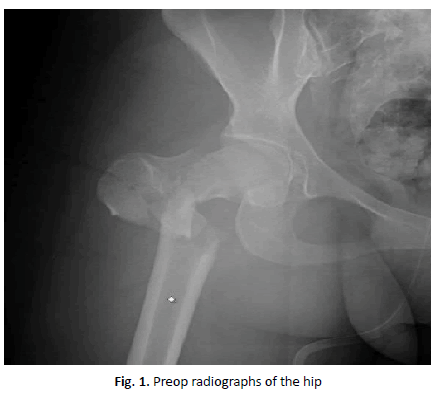
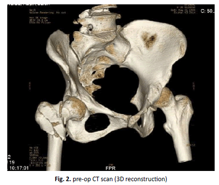
At the time of presentation, the patient was conscious, alert, oriented and hemodynamically stable. The patient had right hip pain and swelling and severe tenderness over the right greater trchanteric area, there was obvious shortening and external rotation of the right lower limb com-pared to the contralateral side. The radiographs (Fig. 1) revealed unstable comminuted pertrochanteric fracture on the right side associated with a partially displaced femoral neck fracture. A noncontrast computed tomography (CT) scan with 3D reconstruction was done (Fig. 2). Which confirmed the two fractures in the right hip.
The patient was admitted and cleared by the anesthesia team and taken to the operating room. The patient and was placed in the left lateral position on a radiolucent surgical table, using a fracture table. The foot of the affected limb was placed in a foot holder to achieve traction and rotation for fracture reduction. The unaffected limb was extended at the hip. The fracture reduction was attempted and checked in both the Anterioposterior (AP) and lateral views with an image intensifier, prior to draping. The femoral neck fracture was carefully assessed for as best a reduction as possible.
A lateral incision was made around 15 cm in length, starting 2 cm above the greater trochanter and extending down. The fascia lata was incised longitudinally. The vastus lateralis was reflected off the proximal femur anteriorly. The anterior one third of gluteus medium was also reflected to expose the fracture. The fracture was identified. The pertrochanteric fracture was comminuted. There was a large fragment of the lesser trochanter that had separated and an extension of the fracture line to the greater trochanter was noted. Further longitudinal traction and the external rotation was applied and fracture reduction assessed with an image intensifier.
Neck of the femur fracture was stabilized first. A guide wire was inserted into the femoral neck, advanced into the femoral head. The position was checked on the AP and lateral view. The femoral neck fracture was stabilized with a 4.5 lag screw.
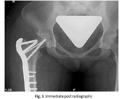
Proximal Femoral Anatomical Plate (PFAP) was used in this case. The large trochanteric plate 5 hole with 3 screws to the femoral head was placed on to the lateral aspect of the proximal femur. The plate locking sleeve was used for the proximal locking screws. The proximal part of the femur and the neck of the femur was fixed first. Proximal screws were placed into the femoral neck and head and 4 distal screws were placed.
Postoperative radiographs showed anatomical reductions of both the femoral neck and the inter-trochanteric fracture (Fig. 3).
Physical therapy was initiated with early mobilization on the same day. Patient was instructed non-weight bearing mobilization for 6 weeks. After 6 weeks of surgery partial weight bearing started and three months after the operation, total weight bearing was allowed. The patient was given enoxaparin 40 mg subcutaneous for 4 weeks post-surgery.
The patient was seen in the clinic two weeks post-surgery for wound assessment and clips removal, the wound healed well.
Laboratory findings, Erythrocyte Sedimentation Ratio (ESR) and C-Reactive Protein (CRP) normalized at 2 weeks post surgery. The patient was followed every three months for the first year and then yearly follow up with radiographs of the hip and pelvis at each visit.
Discussion
Femoral neck fracture is one of the most common fracture, accounting for 50% of hip fractures. The occurrence of femoral neck fracture in young adults is usually caused by high-energy injuries such as traffic injury, fall from the heights. In the elderly it is because of poor bone quality and associated with minor falls. Femoral shaft fractures have been the most common fracture associated with the ipsilateral femoral neck fracture, but a concomitant pertrochanteric fracture was associated rarely [2].
There are twelve cases reported in the medical literature on ipsilateral fractures of the femoral neck and perertrochanteric regions. The majority of the cases were intertrochanteric fractures accompanied with undisplaced femoral neck fractures in the ipsilateral femur in the elderly associated with osteoporosis [1]. Only three cases were reported in young patients [1] who sustained femoral neck fracture accompanied with intertrochanteric fracture and managed with different fixation devices. we had a young patient who had a high energy trauma in a motor vehicle accident. She was a front seat passenger. She sustained pertrochanteric fracture accompanied with neck of femur fracture. This was evident on the preoperative plain radiographs and the CT scan revealed the fracture pattern. Four basic principles were followed to get the best possible outcome. Anatomic reduction, stable fixation, preservation of blood supply, early active mobilization.
Devdatta et al. [1] reported a case of Ipsilateral femoral neck and trochanter fracture managed with a dynamic condylar screw and a cannulated screw, the patient had good result at 1 year. Both the fractures united.
This injury being rarely seen can easily be missed on plain radiographic evaluation. Of the cases reported in the literature, five cases were apparent at initial radiographic evaluation [3-7].
Three were confirmed on further imaging preoperatively [8-10]. two were identified by fluoroscopy during surgical procedure [11,12] while one was identified in a postoperative period. [5] In our case, the AP view did reveal the presence of a fracture line in the femoral neck region. A CT scan with a 3D reconstruction of the pelvis performed for the preoperative evaluation of the fracture revealed a fracture line at the femoral neck. Thus, a CT scan with 3D reformatting is very helpful not only in the preoperative diagnosis of the fracture but it also defines the fracture pattern to plan the surgery.
We chose PFAP because it has a lot of factors that will help to have a good outcome. PFAP is an atomically contoured to approximate on the lateral aspect of the proximal femur and these plates specifically designed for left or right femurs to accommodate average femoral neck anteversion. Plate lengths allow spanning of the entire diaphysis in segmental fracture patterns. Use of the locking screws provides the option of applying screws in different angles and direction to provide a stable construct independent of bone quality. Plates can be tensioned to create a load-sharing construct plate. It has three proximal screw holes. First proximal hole (7.3 mm), 95°, second proximal hole (7.3 mm), 120° and third proximal hole (5.0 mm), 135°. In reported cases there is no case reported to manage the femoral neck and pertrochanteric fractures with the PFAP.
Six of the reported cases were fixed with DHS, successful result was present in five cases while one case [13] in whom fracture was recognized postoperatively had fixation failure. One case [1] with pinning in situ died from complications not related to surgery. Good result was also seen with hemiarthroplasty [10,11]. However arthroplasty is reserved for the elderly with poor bone stock and sedentary life style.
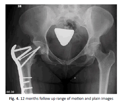
At 4-month follow-up, the case who had a fixation with? PCCP [4] had a good result. The final case with cancellous cannulated screws, Knowles pin, and DCP [7] also had good result at 1 year. We follow up patient for five years. At this time the patient was pain frees with no limitations of activities. He was back to his recreational and sporting activities and had achieved then premorbid status. Both the fractures had united with no signs of avascular necrosis or arthritis (Fig. 4).
Conclusion
The concomitant displaced neck and intertrochanteric fractures in the ipsilateral femur in a young patient is very rare. We managed this patient successfully with a PAFP with excellent clinical and radiographic outcome at five years post-surgery.
REFERENCES
- Devdatta S.N., Kumar K.V., Trikha V., et al.: Ipsilateral femoral neck and trochanter fracture. Indian J Orthop. 2011;45:82-86.
- Seyegh F., Karataglis D., Trapotsis S., et al.: Concomitant ipsilateral pertrochanteric and subcapital fracture of the proximal femur. Eur J Trauma. 2005;31:64-67.
- Kumar R., Khan R., Moholkar K., et al.: A rare combination fracture of the neck of femur. Eur J Orthop Surg Traumatol. 2001;11:59-61.
- Poulter R.J., Ashworth M.J.: Concomitant ipsilateral subcapital and intertrochanteric fractures of the femur. Injury Ext. 2007;38:88-89.
- Sayegh F., Karataglis D., Trapotsis S., et al.: Concomitant ipsilateral pertro-chanteric and subcapital fracture of the proximal femur. Eur J Trauma. 2005;31:64-67.
- Butt M.F., Dhar S.A., Hussain A., et al.: Femoral neck fracture with ipsilateral trochanteric fracture: Is there room for osteosynthesis? Internet J Orthop Surg. 2007;22:5.
- Dhar S.A., Mir M.R., Butt M.F., et al.: Osteosynthesis for a T-shaped fracture of the femoral neck and trochanter: A case report. J Orthop Surg. 2008;16:257-259.
- Lawrence B., Isaacs C.: Concomitant ipsilateral intertrochanteric and subcapital fracture of the hip. J Orthop Trauma. 1993;7:146-148.
- Pemberton D.J., Kriebich D.N., Moran C.G.: Segmental fracture of the neck of the femur. Injury. 1989;20:306.
- Yuzo O., Yamanaka M., Hiroshi T., et al.: A case of femoral neck and trochanteric fracture in ipsilateral femur. Orthop Traumatol. 2001;50:1072-1075.
- An H.S., Wojcieszek J.M., Cooke R.F., et al.: Simultaneous ipsilateral intertrochanteric and subcapital fracture of the hip. A case report. Orthopedics. 1989;2:721-723.
- Cohen I., Rzetelny V.: Simultaneous ipsilateral pertrochanteric and subcapital fractures. Orthopaedics. 1999;22:535-536.
- Perry D.C., Scott S.J.: Concomitant ipsilateral intracapsular and extracapsular femoral neck fracture: a case report. J Med Case Reports. 2008;2:68-70.


 Journal of Orthopaedics Trauma Surgery and Related Research a publication of Polish Society, is a peer-reviewed online journal with quaterly print on demand compilation of issues published.