Effect of visceral mobilization on vital capacity in scoliosis among adolescent females
2 Department of Physiotherapy, United college off Physical Therapy, Karachi, Pakistan, Email: rashadabdul66@gmail.com
3 Department of Physiotherapy, Pakistan Institute of Rehabilitation and medical Sciences, Karachi, Pakistan, Email: Rabiaintekhab22@aol.com
4 Department of Physiotherapy, Mamji Hospital, Karachi, Pakistan, Email: abc@gmail.com
5 Department of Physiotherapy, Dr Essa Physiotherapy & Rehabilitation Center, Karachi, Pakistan, Email: abc@gmail.com
6 Department of Physiotherapy, Dr Essa Physiotherapy & Rehabilitation Center, Karachi, Pakistan, Email: Alishanoreen2@yahoo.com
7 Department of Physiotherapy, General Creek Hospital, Karachi, Pakistan, Email: abc@gmail.com
Received: 05-Jan-2021 Accepted Date: Jan 19, 2021 ; Published: 02-Feb-2021, DOI: 10.37532/1897-2276.2021.16(1).4
This open-access article is distributed under the terms of the Creative Commons Attribution Non-Commercial License (CC BY-NC) (http://creativecommons.org/licenses/by-nc/4.0/), which permits reuse, distribution and reproduction of the article, provided that the original work is properly cited and the reuse is restricted to noncommercial purposes. For commercial reuse, contact reprints@pulsus.com
Abstract
Background: Scoliosis is lateral deviation of spine mostly on loading area of spine. Idiopathic Scoliosis (IS), very common form of scoliosis which cause is unknown but some triggering events gives believe to leads this and resulting in spinal deformation in spinal curvature (measured in cobb’s angle) and irregular loading of spine. Objective: To determine the effects of visceral mobilization on vital capacity and Cobb’s angle in scoliosis among adolescent females. Methods: Total nine female patients were participated in the study. Age ranges from 9-11 years with idiopathic spine scoliosis, Cobb’s angle 100 or above were included. Treatment was given including conventional physical therapy followed by visceral mobilization techniques for chest and associated muscles of respiration. Treatment time was 45-60 minutes and total 18 sessions were given in 6 weeks with the frequency of 3 sessions per week. Result: The overall mean age of the participants was 16.77 ± 4.95 years. Statistically significant results were also obtained in Force Vital Capacity with the p-value of 0.05 and there was significant improvements in the Cobb’s angle of the participants were noticed with the p-value of 0.02. Conclusion: Visceral mobilization has a significant impact on Force Vital Capacity (FVC) and eventually in reduction of Cobb’s angle in scoliotic patients.
Keywords
respiration, spine, scoliosis, spinal curvature, vital capacity
Introduction
Scoliosis is a deviation of the normal vertical line of the spine, consisting of a lateral curvature with the rotation of the vertebrae within the curve [1]. It can be characterized into three types-idiopathic, congenital and syndromic [2]. Idiopathic Scoliosis (IS), a very common form of scoliosis which causes is unknown but some triggering events give believe to leads to this and resulting in spinal deformation in spinal curvature (measured in cobb’s angle) and irregular loading of the spine [3]. Cobb Angle is considerably greater in females than in males: The female to male ratio rises from 1.4:1 in curves from 10 to 20 degrees up to 7.2:1 in curves greater than 40 degrees [4]. Pulmonary complications are common in Scoliosis patients, but in opposite relation to spinal curves, which means as Cobbs angle increases there is a substantial decrease found in respiratory function [5]. Impaired respiratory function is a common problem of adults with idiopathic scoliosis, because of Ribcage and spine deformity. Many factors that can affect respiratory functions in patients with idiopathic scoliosis mainly identified are structural dysfunction, thoracic curvature, apex displacement, hypo kyphosis, or lordosis of the thoracic spine, and the vertebras involved in the main curve [6]. The normal mechanism of ventilation depends on the rib cage compliance and mobility, but in association with spinal deformity, the normal ventilatory function becomes altered and led to the decreased capability to do physical activities [7]. In patients with scoliosis, there is a decrease in inspiratory muscle forces that results in insufficient mechanical coupling of ribcage and muscles of inspiration [8]. The primary goal of rehabilitation for AIS (Adolescence Idiopathic Scoliosis) is to reduce the progression of curves thereby decreasing the risk of secondary impairment, including back pain, breathing problems, cosmetic deformities, and improve the quality of life and also to correct alignment of trunk muscles, stimulation of movement of the trunk during and also improves pulmonary functions. Traditional treatment includes exercises, Milwaukee or Boston orthosis, Electrical stimulation, and extension therapy [9].
Another treatment option for idiopathic scoliosis is Manual therapy which includes thrust mobilization techniques, spinal distraction, soft tissue techniques, craniosacral therapy, visceral mobilization or manipulation, and other techniques [10]. Different manual therapy techniques were presented to improve respiratory functions that targeted lymphatic, neuronal, and musculoskeletal components of the respiratory system [11]. Increasing the posterior-anterior chest dimension that aids to normalize the pulmonary mechanics, those dependents on amplitude of ribs movements and cost vertebral joint axis [12].
Chest mobilization assists to improve chest wall flexibility and thoracic spine mobility. The mechanism comprises the lengthening of intercostal muscles and consequently helps in improving muscle performance. This increases biomechanics of chest mobility by improving the direction of anterior upward of upper coastal and lateral outward of lower coastal movement, that including downward of diaphragm directions. Increasing chest mobility with improvement in respiratory muscle contraction can assist in increasing lung volume, coughing efficiency, and breathing control [13]. Past researches have investigated the procedure of thoracic joint activation and thoracic mobility exercises to improve deformations of the vertebrae or chest and therefore upgrade respiratory function. These interventions improved pulmonary capacity and thoracic flexibility and improved respiratory muscle work, chest development, and diaphragm movement by decreasing the stiffness of tissues and intervertebral discs by improving the endurance and vertebra extensor muscles stretch thoracic flexibility training [14]. The conventional, evidence-based medicine in the administration of scoliosis has acquired tremendous contribution under the expression “Physiotherapy Scoliosis Specific Activities” (PSSE) instituted by “the Society of Scoliosis Orthopedic Rehabilitation and Treatment” (SOSORT). From that point forward various methodologies have been created under PSSE that asserts the adequacy of recently created practice systems in the administration of Cobb’s angle, functional capacity among patients with idiopathic scoliosis [15]. The purpose of this study was to see the effect of visceral mobilization on vital capacity in scoliosis among adolescent females.
Methods
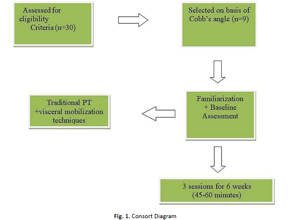
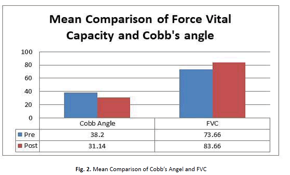
It was a pre and post-experimental study conducted from January 2018 to March 2019 in the outpatient department of Physical therapy and Rehabilitation, Ziauddin University. The sampling technique was simple random sampling. The ethical review committee of the Ziauddin University Clifton campus approved to conduct of this study. The Source of funding for this study is self so absolutely hasn’t any influence on this paper. The participants in the study were first screened by using Adam’s forward bend test, with positive results of Adam’s forward bend test participants were ask for the PA X-ray of the thoracic spine. Based on Cobb’s angle individuals were recruited for the study. Treatment was given for six weeks including traditional physical therapy (Hot packs, Cold packs, Electrotherapy) followed by visceral mobilization techniques for the chest and associated muscles of respiration. Exercises were included mobilization of respiratory muscles along with visceral mobilization.(Fig 1 and 2).
Adam’s Forward Bend Test
A patient takes off his/her t-shirt so that the spine is visible. The patient needs to bend forward, starting at the waist until the back comes in the horizontal plane, with the feet together, arms hanging, and the knees in extension. The palms are held together. The examiner stands at the back of the patient and looks along the horizontal plane of the spine, searching for abnormalities of the spinal curve, like increased or decreased lordosis/kyphosis, and an asymmetry of the trunk [16].(Table 1)
| Variable | Pre Mean ± SD | Post Mean ± SD | p-value |
|---|---|---|---|
| FVC | 73.66 ± 10.03 | 83.66 ± 23.14 | 0.05 |
| Cobb’s Angle | 38.22 ± 18.97 | 31.0 ± 14.34 | 0.02 |
Table 1 Mean Comparison of Force Vital Capacity.
Cobb’s Angle
Cobb Angle is used as a standard measurement to determine and track the progression of scoliosis. The measurement of the Cobb angle involves estimating the angle between the two tangents of the upper and lower endplates of the upper and lower end vertebra, respectively [17].
Spirometer
Spirometry is a physiological test for assessing the functional aspect of the lungs using an objective indicator by measuring the amount of air that a patient can inhale and exhale to the maximum. Subjects undergoing spirometry are asked to inhale the air quickly up to a Total Lung Capacity (TLC) at once. Subjects are then asked to exhale as hard as possible until they cannot exhale any longer. Thereafter, they inhale immediately as rapidly as possible [18].
Treatment for ligaments of the Coracoid Process
Check the trapezoid, coracoacromial, and conoid ligaments for compassion. To take care of, apply resistances to the perceptive parts until the soreness has vanished. Force on the perceptive parts should be tough sufficient to hardly cross the pain. Treatment success can then be assessed adequately.
Compression and Decompression of the Clavicle along the longitudinal Axis
Grasp the clavicle-Acromial end, between the thenar and hypothenar. Then with medial handgrip the clavicle sternal end, similarly. Place fingers of both hands over each other over the clavicle.
Fascial Mobilization of the Clavicle
Set the index phalanges of two hands on the back area of the clavicle Such that it feels along in its whole length. Then position the thumbs of the two hands in like manner on the front surface.
Compression and Decompression of the Sternum
Through the cranial hand, grip the cranial end of the sternum between thenar and hypo-thenar. Through the caudal hand, hold the xiphoid end of the sternum in the same way. Position the fingers of the two hands over each other on the sternum.
Movement of the Corpomanubrial Intersection of the Sternum
Position cranial hand at manubrium with the thenar next to the intersection point on the sternal body. The caudal hand should lies on the sternal body and thenar on the edge of the manubrium. The intersection among manubrium and sternal body should now lie precisely between the two thenars.
Moving upward of the Corpoxiphoid intersection of the Sternum
Position your cranial hand on top of the sternal body with the thenar at the intersection of the xiphoid procedure. The caudal hand should lie on the xiphoid and thenar on the edge to the sternal body. The intersection amongst xiphoid and sterna body now lies precisely sandwiched between the two thenars.
Movement of the Transversus Thoracic
Assume the transversus thoracic as a top to bottom Christmas tree. Position your caudal hand on the inferior third of the sternum and the cranial hand crosswise the other hand, contra sideways to the costochondral intersection on the ribs (on the left, ribs 2-5; on the right, ribs 3-6).
Pectoral lift
With a squeeze hold, get a handle on the Pectoralis major and minor on the two sides with your hands. With a solid grasp, drag the tissue cranially while maintaining the position for up to 2 min. You will rapidly notice a clear fascial let loose. If the drag is hurting at the start, this pain will vanish after only a minimum period.
Movement of the Mediastinum
Make a position for the front hand with the fingertips pointing cranial lie on the lower one-third of the patient’s sternum. The dorsal surface of the hand likewise lies with the fingertips pointing cranially on the spinal section at the level of the manubrium sterni (Hebgen 1011).
The frequency of each mobilization is 3-5 times in a single session. The treatment time was 45 minutes - 60 minutes. A total of 18 sessions were conducted in 6 weeks with the frequency of 3 sessions per week. The pre and post-assessment of Cobb’s angle and Spirometer reading of the participants recruited in this study was done on the first day before the exercise session and after the completion of the six weeks of the exercise protocol. Data collection was done by spirolab before the start of the treatment session and after the last session. Data were stored and analyzed using SPSS version 20.0.
Results
A total of 30 patients were assessed for scoliosis; only 9 patients met the inclusion criteria and were included in the study. The mean age of participants was 16 years and BMI was 19. Paired sample t-test was used to see the mean differences of Forced Vital Capacity and Cobb’s Angle at pre and post-level of treatment. Meanwhile statistically significant results were also obtained in FVC with the p-value of 0.05 and there were significant improvements in Cobb’s angle of the participants were noticed with the p-value of 0.02, after the completion of the exercise regime. As reported in the Table 1.
Discussion
This study was conducted to determine the effects of visceral mobilization on vital capacity in scoliosis among adolescent females. The results showed a significant decrease observed in Cobb’s angle after the treatment with a p-value of 0.022. After the treatment, FVC gets better as compared to pretreatment. However, this difference was statistically significant with a p-value of 0.05. A study was performed by Sungyoung Yoon et al 2016 to check specific exercises of physical therapy scoliosis on idiopathic scoliosis patients. A 15-years old middle school male student with scoliosis. He was measured before and after 8 weeks of training for breathing pattern, Cobb’s angle, and angle of rotation of the spine. After training of 8 weeks the breathing pattern, Cobb’s angle, and angle of rotation of spin were improved [19].
A randomized control trial was conducted by Abdelaal Ashraf et al. 2015 to study the effect of coastal and diaphragmatic manipulation on functional capacity and respiratory function in COPD patients, he reported that Diaphragmatic manipulation procedures produced substantial benefits on both functional capacity and pulmonary function in COPD. Moreover; outcome shows better responses in combine the application of intervention in pulmonary function and functional capacity [11]. A study conducted by Jae-Man Yang et al. reported that combined application of strengthening exercises, Schroth, and stretching may help decrease Cobb’s angle and the rib bulge in idiopathic scoliosis [20]. Carlos Cruz-Montecinos et al reported that the soft tissue protocol of manual therapy appears to have the possible immediate clinically significant improvement in pulmonary function [21]. This study indicates the focus on visceral mobilization and the impact of visceral mobilization to reduce Cobb’s angle in scoliotic patients. Studies show a significant reduction of Cobb’s angle, in scoliotic patients. Therefore, it’s essential to check the conventional approaches to make sure the foremost effective approaches are used. Especially when considering that in the time of life growth spurt there’s no time to waste, from the patients’ viewpoint, with unverified strategies of treatment.
Conclusion
Visceral mobilization has a significant impact on Force Vital Capacity (FVC) and eventually in the reduction of Cobb’s angle in scoliotic patients.
Limitations
This study was done on a small population due to a limited number of patients. There was no control group. Due to the small sample size, we lack to establish a strong correlation. Besides this our population was only females and belongs to a good socioeconomic class, future studies are needed to be conducted on both gender and large ethnic group to establish the generalizability of the treatment protocol.
REFERENCES
- Choudhry M.N., Ahmad Z., Verma R.: Adolescent idiopathic scoliosis. Open Ortho J. 2016;10:143.
- Altaf F., Gibson A., Dannawi Z., et al.: Adolescent idiopathic scoliosis. Bmj. 2013;30:346.
- Lebel A., Lebel V.A.: Severe progressive scoliosis in an adult female possibly secondary thoracic surgery in childhood treated with scoliosis specific Schroth physiotherapy: Case presentation. Scoliosis and spinal disorders. 2016;11:41.
- Etemadifar M., Hadi A., Nazem K., et al.: Epidemiology of adolescent idiopathic scoliosis in Isfahan, Iran: A school-based study during 2014-2015. J Res Med Sci. 2020;25:48.
- Chun E.M., Suh S.W., Modi H.N., et al.: The change in ratio of convex and concave lung volume in adolescent idiopathic scoliosis: a 3D CT scan based cross sectional study of effect of severity of curve on convex and concave lung volumes in 99 cases. Euro Spine J. 2008;17:224-229.
- Fujita N., Yagi M., Michikawa T., et al: Impact of fusion for adolescent idiopathic scoliosis on lung volume measured with computed tomography. Euro Spine J. 2019;28:2034-2041.
- Sperandio E.F., Vidotto M.C., Alexandre A.S., et al.: Functional exercise capacity, lung function and chest wall deformity in patients with adolescent idiopathic scoliosis. Fisioterapia em Movimento. 2015;28:563-572.
- Szeinberg A., Canny G.J., Rashed N.,et al.: Forced vital capacity and maximal respiratory pressures in patients with mild and moderate scoliosis. Pediatric Pulmonol. 1988;4:8-12.
- Kim J.J., Song G.B., Park E.C.: Effects of Swiss ball exercise and resistance exercise on respiratory function and trunk control ability in patients with scoliosis. J Phy Therapy Sci. 2015;27:1775-1778.
- Jung G.H., Lee H., Kong H.J., et al.: A Systematic review of chuna manual therapy for adolescent idiopathic scoliosis. J Acupuncture Res. 2019;36:119-130.
- Abdelaal A.A., Ali M.M., Hegazy I.M.: Effect of diaphragmatic and costal manipulation on pulmonary function and functional capacity in chronic obstructive pulmonary disease patients: Randomized controlled study. Int J Med Res Health Sci. 2015;4:841-847.
- Wnuk B., Blicharska I., Blaszczak E., et al.: The impact of the derotational mobilization of manual therapy according to kaltenborn-evjenth on the angle of trunk rotation in patients with adolescent idiopathic scoliosis--pilot study. Ortopedia Traumatologia Rehabilitacja. 2015;17:343-350.
- Ong K.C.: Chronic Obstructive Pulmonary Disease: Current Concepts and Practice. BoD-Books on Demand.2012.
- Jung J.H., Moon D.C.: The effect of thoracic region self-mobilization on chest expansion and pulmonary function. J Phy Ther Sci. 2015;27:2779-2781.
- Farooqui S.I., Siddiqui PQ., Basit Ansari AF.: Effects of spinal mobilization techniques in the management of adolescent idiopathic scoliosis-A meta-analysis. Int J Health Sci. 2018;12:44.
- Reamy V.B.: Adolescent idiopathic scoliosis: Review and current concepts. American Family Physician. 2001;1:1
- Horng H.M., Pang K.C., Jun F.M., et al.: Cobb Angle Measurement of Spine from X-Ray Images using convolutional neural network. Computational and Mathematical Methods in Medicine. 2019;19:1-18
- Sim S.Y., Hyun L.J., Yeon L.W., et al.: Spirometry and Bronchodilator Test. Tuberc Respir Dis. 2017;80:105-112
- Yoon S., Rhee M.H.: Effect of physical therapy scoliosis specific exercises using breathing pattern on adolescent idiopathic scoliosis. J Phy Ther Sci. 2016;28:3261-3263.
- Yang J.M., Lee J.H., Lee D.H.: Effects of consecutive application of stretching, Schroth, and strengthening exercises on Cobb’s angle and the rib hump in an adult with idiopathic scoliosis. J Phy Ther Sci. 2015;27:2667-2669.
- Cruz-Montecinos C., Godoy-Olave D., Contreras-Briceño F.A., et al.: The immediate effect of soft tissue manual therapy intervention on lung function in severe chronic obstructive pulmonary disease. Int J Chronic Obstructive Pulmonary Disease. 2017;12:691


 Journal of Orthopaedics Trauma Surgery and Related Research a publication of Polish Society, is a peer-reviewed online journal with quaterly print on demand compilation of issues published.