Does flexion contracture really matter in medial open wedge high tibial osteotomy?
Received: 22-Nov-2018 Accepted Date: Dec 27, 2018 ; Published: 03-Jan-2019
This open-access article is distributed under the terms of the Creative Commons Attribution Non-Commercial License (CC BY-NC) (http://creativecommons.org/licenses/by-nc/4.0/), which permits reuse, distribution and reproduction of the article, provided that the original work is properly cited and the reuse is restricted to noncommercial purposes. For commercial reuse, contact reprints@pulsus.com
Abstract
Objective: Several studies stated on medial open wedge tibial osteotomy but there is still some debate about the acceptable amount of pre-operative flexion contracture degree. Also, clinical effects of alteration of tibial slope after the procedure is not clear. This study aimed to investigate the mid-term clinical and radiological findings and complications of medial open wedge tibial osteotomy. Materials and methods: 44 knees of 42 patients were investigated retrospectively between January 2001 and February 2012. Tibial sagittal slope, mechanical tibiofemoral angle (mTFA), mechanical lateral distal femoral angle (mLDFA) and Medial Proximal Tibia Angle (MPTA) were measured, pre and post-operatively. Clinical outcome was evaluated with HSS (Hospital for Special Surgery) Knee Score, OKS (Oxford Knee Score) and KOSADLS (Knee Outcome Survey-Activities of Daily Living Scale). Results: The mean age of the participants was 45.7 ± 18.3 (range, 17-84), and the mean follow-up was 92 ± 7 (range, 70-113) months. Gender distrubition was 34 (%81) female and 8 (%19) male. 10 degrees of flexion contracture was present in 4 (%10) patients preoperatively. Mean knee range of motion was increased from 120 ± 11 to 130 ± 9 degrees, postoperatively. HSS scores were improved to excellent in 29 (69%), good in 9 (21%) and moderate in 4 (10%). ADLS and Oxford scores were improved two-fold. Conclusion: Ten degrees of flexion contracture may not be a restraint for osteotomy, with the use of technical details to prevent slope increase. Even with the rise of the slope, up to 10 degrees of flexion contracture could be corrected without anterior wedge resection or posterior capsular release. Level of evidence: Level IV, Case series, case control study (diagnostic studies), poor reference standard, analyses with no sensitivity analyses.
Keywords
High, tibia, osteotomy, open, wedge, flexion, contracture
Introduction
Medial gonarthrosis causes knee pain and restriction of daily activities in elderly [1]. High tibial osteotomy is more appropriate procedure than arthroplasty in young patients with early osteoarthritis [2]. Lower extremity amechanical axis could be improved and total knee replacement in future could be avoided by high tibial osteotomy [3]. Various tibial osteotomy types (e.g., closing wedge, opening wedge, dome osteotomy) were defined in this purpose. Osteotomy can also be added to osteochondroplasty, menisectomy or instability procedures aiming to facilitate cartilage preservation, subchondral healing and to increase stability [4]. To our knowledge, several studies investigate medial open wedge tibial osteotomy, but there is still some debate about the acceptable amount of pre-operative flexion contracture degree. Also, clinical effects of alteration of tibial slope after the procedure is not clear. This study aimed to evaluate the clinical effects of pre-operative flexion contracture degree and alteration of tibial slope. Also, we investigated the mid-term clinical and radiological results and complications of medial open-wedge tibial osteotomy.
Materials and Methods
Institutional review board approval and informed consent was obtained from the patients. Both the Belmont report on ethical principles and the National Institute of Health (NIH) guidelines were considered in this study. Between January 2001 and February 2012, a total of 54 knees of 52 patients with high tibial osteotomy were investigated retrospectively. Inclusion criteria were medial knee pain, isolated medial compartment osteoarthritis or osteonecrosis of the medial compartment, malalignment with 5-15 degrees varus between the tibial and femoral mechanical axis, medial open-wedge osteotomy and fixation with a wedge plate. Anterior cruciate ligament insufficiency, symptomatic osteoarthrosis of the lateral or patellofemoral compartment, osteotomy added to osteochondroplasty, menisectomy, instability surgery, and revision cases were excluded. Five patients were died and five patients were lost to follow-up. After eligibility criteria 44 knees of 42 patients were included in the study. Knee ROM (Range of Motion), instability, contracture and muscle strength were assessed by physical evaluation. Anteroposterior, lateral, tangential X-rays and leg length orthoroentgenogram while standing were obtained for both of the lower extremity for all patients. Ahlback classification was used to evaluate the osteoarthritis for operation [1]. Two different surgeons performed the operations. A longitudinal incision is extending to 7 cm distal of the joint, between the tibial tuberosity and MCL (Medial Collateral Ligament) was made. The anterior fibers of the superficial MCL were cut, and a retractor was placed to the posteromedial corner. A medial open-wedge osteotomy was performed and fixed with a wedge plate to establish the needed correction angle. 1 g cephalosporin was administered prior to skin incision. On the first post-operative day, low-molecule weight heparin was applied subcutaneously and for 1 month. Patients were allowed to sit and perform isometric quadriceps exercises. On the second day, drains were taken, and patients were allowed to walk with no load. Weight-bearing was allowed at post-operative 6th week. All patients were followed up at 6, 12, 18 and 24 weeks and after that six months until the last review. All patients were assessed with anterior-posterior and lateral X-rays while standing. Mechanical axis, femorotibial angle were measured and compared with the preoperative values. Tibial slope angle was measured by using tibial anatomic axis. Knee ROM, HSS (Hospital for Special Surgery) Knee Score, KOS-ADLS (Knee Outcome Survey- Activities of Daily Living Scale) and OKS (Oxford Knee Score) were used to evaluate the clinical results.
Accepting less than 5% probability of a type I error and a power of 80%, the required sample size was 34. SSPS version 11 (SPSS, Inc., Chicago, IL, USA) was used for statistical analysis, and data were presented as the mean and standard deviations. Postoperative functional results and osteoarthrosis radiological classification data were compared using Ficher’s exact test.
Preoperative and postoperative data of the scoring systems used in the study were compared with Student’s t-test. A value of p<0.05 was considered statistically significant.
Results
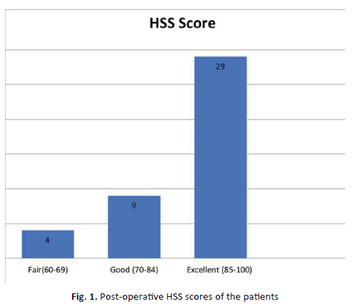
The mean age of the participants was 45.7 ± 18.3 (range, 17-84), and the mean follow-up was 92 ± 7 (range, 70-113) months. The mean body mass index was 32 ± 2 kg/m2. According to Ahlback criteria (5), 23 (55%) and 19 (44%) patients had stage 1 and stage 2 arthrosis, respectively. The mean mechanical axis was 7.8 ± 3.04 degrees varus preoperatively and 0.8 ± 3.32 degrees valgus postoperatively. The mean anatomic axis was 5.7 ± 2.22 degrees varus preoperatively and 2.7 ± 3.46 degrees valgus postoperatively. Also, the mean knee range of motion increased 10 ± 10 degrees postoperatively. Flexion contracture was present in 4 patients (up to 10 degrees) and improved to 0 degrees in all, postoperatively. The measured HSS scores were excellent to good in 38 (90%) patients (Fig. 1). The ADLS and Oxford scores improved from 35.83 ± 2.8 and 42.58 ± 4.5 to 71.24 ± 5.2 and 21.62 ± 4.4, respectively (Table 1). Mean tibial slope angle was increased to 2.34 ± 1,18 degrees after surgery. Among the participants, 22 (52%) were very satisfied, 13 (31%) were satisfied, 6 (14%) were moderately and 1 (3%) was mildly satisfied with the results of the surgery. A tibia nondisplaced lateral plateau fracture was encountered in one patient and treated with long leg brace. There was a superficial wound problem in one patient and was successfully treated with oral antibiotics. Both of them did not require secondary intervention. No implant insufficiency, deep venous thrombosis or pulmonary embolism were encountered.
| Preoperative | Postoperative | p value* | |
|---|---|---|---|
| OKS | 42.58±4.5 | 21.62±4.4 | p=0.000032 |
| KOS-ADSL | 35.83±2.8 | 71.24±5.2 | p=0.000073 |
| HSS Knee Score | 65±3.27 | 82±3.82 | P=0.000045 |
| Knee ROM | 120±11 | 130±9 | p=0.000034 |
| Mechnaical Axis | 7.8±3.04 varus |
0.8±3.32 valgus |
p=0.000053 |
| Anatomical Axis | 5.7±2.22 varus |
2.7±3.46 valgus |
p=0.000071 |
Table 1: Post-operative HSS scores of the patients
Discussion
Medial opening wedge high tibial osteotomy has become more popular than lateral closing wedge osteotomy. Generally tibial slope increases after open-wedge and decreases after closing-wedge high tibial osteotomy [5]. It has been recommended that the osteotomy line in the sagittal plane be parallel to the medial posterior tibial slope [6]. However, the effects on posterior tibial slope of closing-or opening-wedge osteotomies remain controversial. The distinctive result of this study was up to 10 degrees of flexion contracture did not restrain the efficacy of tibial open-wedge osteotomy. However, the participants did not exhibit anterior wedge resection or posterior capsule release.
There are some reports that high tibial osteotomy has no effect on the range of motion, and over 5 degrees of flexion contracture signifies contraindication for osteotomy [1,7,8]. Naudie et al., found that a preoperative ROM lower than 120° associated with flexion contracture greater than 5° was related to early failure (p-value 0.042) [7]. Flexion contracture as a contraindication is based on relatively poor results of small epidemiological studies. Deflexion effect can be achieved by reducing the posterior slope in cases with flexion contracture. But this may lead to anterior translation and an increased load on the anterior cruciate ligament. To our knowledge there is only one study focuses on preoperative flexion contracture and reported satisfactory results with severe (>20 degrees) flexion contracture [9].
Ducat et al., suggested loosening soft tissue and performing an osteotomy in the posterior to avoid slope increase. But, osteotomy may result with recurvation with anterior wedge resection or posterior capsulotomy [10-12]. Noyes et al., reported that anterior osteotomy gap should be half as large as the posteromedial gap to obtain a standard posterior tibial slope [13]. Shi et al., found that a 1 degree increase in the posterior tibial slope resulted in a 1.8 degree increase in knee flexion [14]. Similarly in our series mean flexion degree increase was 10 degrees but mean slope increase was only 2, 34 degrees. This may be related to the disappearance of the protective muscle spasm due to the newly formed load distribution [15].
According to a meta-analysis of sex differences in osteoarthritis, females who <55 years tended to have more severe OA in the knee. These results demonstrate the presence of sex differences in OA prevalence and incidence. Females also tend to have more severe knee OA, particularly after reaching menopausal age [16]. Van Houten et al., reported a sex ratio of 3:1, BMI of 28 ± 4 kg/m2 and complication rates of 17% in 192 patients and 224 knees [17], while Goshima et al., reported a sex ratio of 1:2, BMI of 24 ± 2.6 kg/m2 and complication rates of 20% in 50 patients and 60 knees [5].
This study was a retrospective and non-comparative study. Prospective study design may provide further information. Also, patellofemoral arthrosis degree and correction variances (e.g., overcorrection, under correction, loss of correction) could be compared with clinical results.
In conclusion, even with slope increases, without anterior wedge resection or posterior capsular release, up to 10 degrees of flexion contracture could be corrected. Therefore, in selected patients, flexion contracture may not be a restraint for osteotomy, especially if the slope increase is prevented.
Conclusion
Ten degrees of flexion contracture may not be a restraint for osteotomy, with the use of technical details to prevent slope increase. Even with the rise of the slope; up to 10 degrees of flexion contracture could be corrected without anterior wedge resection or posterior capsular release.
Acknowledgement
None
REFERENCES
- Ahlback S.: Osteoarthrosis of the knee. A radiographic investigation. Acta Radiol Diagn (Stockh). 1968;277:7-72.
- Aydogdu S.: Yuksek Tibial Osteotomi: Sonuclar-Uzun Donem Sonuclar [High Tibial Osteotomy-Long Term Results]. In: Sur H (ed) Yuksek Tibial Osteotomi [High Tibial Osteotomy]. Ankara: TOTBID Yayinlari. 2014:97.
- Bombaci H., Canbora K., Onur G., et al.: The effect of open wedge osteotomy on the posterior tibial slope. [Article in Turkish] Acta Orthop Traumatol Turc. 2005;39:404-410.
- Coventry M.B.: Osteotomy about the knee for degenerative and rheumatoid arthritis. J Bone Joint Surg. 1973;55(1):23-48.
- Nha K.W., Kim H.J., Ahn H.S., et al.: Change in posterior tibial slope after open-wedge and closed-wedge high tibial osteotomy: A meta-analysis. Am J Sports Med. 2016;44:3006-3013.
- Lee S.Y., Lim H.C., Bae J.H., et al.: Sagittal osteotomy inclination in medial open-wedge high tibial osteotomy. Knee Surg Sports Traumatol Arthrosc. 2017;25:823-831.
- Naudie D., Bourne R.B., Rorabeck C.H., et al.: The install award. survivorship of the high tibial valgus osteotomy. A 10-to-22-year follow-up study. Clin Orthop Relat Res. 1999:18-27.
- Goshima K., Sawaguchi T., Sakagoshi D., et al.: Age does not affect the clinical and radiological outcomes after open-wedge high tibial osteotomy. Knee Surg Sports Traumatol Arthrosc. 2017;25:918-923.
- Takahashi A.: Clinical results after high tibial osteotomy for medial compartmental osteoarthritis of the knee with flexion contracture above 20°. Japanese Journal of Rheumatism and Joint Surgery. 1991;10:455-462.
- Hassanin A.M., El-Husseiny E.H.M., et al.: Evidence-based medicine in high tibial osteotomy for knee osteoarthritis. Benha Med J. 2015;32:87-91.
- Hernigou P., Medevielle D., Debeyre J., et al.: Proximal tibial osteotomy for osteoarthritis with varus deformity. A ten to thirteen-year follow-up study. J Bone Joint Surg Am. 1987;69:332-354.
- Ducat A., Sariali E., Lebel B., et al.: Posterior tibial slope changes after opening- and closing-wedge high tibial osteotomy: A comparative prospective multicenter study. Orthop Traumatol Surg Res. 2012;98:68-74.
- Noyes F.R., Goebel S.X., West J.: Opening wedge tibial osteotomy: The 3-triangle method to correct axial alignment and tibial slope. Am J Sports Med. 2005;33:378-387.
- Shi X., Shen B., Kang P., et al.: The effect of posterior tibial slope on knee flexion in posterior-stabilized total knee arthroplasty. Knee Surg Sports Traumatol Arthrosc. 2013;21:2696-2703.
- Magyar G., Toksvig-Larsen S., Lindstrand A.: Changes in osseous correction after proximal tibial osteotomy: Radiostereometry of closed- and open-wedge osteotomy in 33 patients. Acta Orthop Scand. 1999;70:473-477.
- Srikanth V.K., Fryer J.L., Zhai G., et al.: A meta-analysis of sex differences prevalence, incidence and severity of osteoarthritis. Osteoarthritis Cartilage. 2005;13:769-781.
- Van-Houten A.H., Heesterbeek P.J., van-Heerwaarden R.J., et al.: Medial open wedge high tibial osteotomy: can delayed or nonunion be predicted? Clin Orthop Relat Res. 2014;472:1217-1223.


 Journal of Orthopaedics Trauma Surgery and Related Research a publication of Polish Society, is a peer-reviewed online journal with quaterly print on demand compilation of issues published.