CT Assessment of accuracy of lumbar pedicle screw insertion (An applied comparative evaluation of conventional and percutaneous techniques)
2 Lecturer of Neurosurgery, Faculty of Medicine, Cairo University, Egypt
3 Neurosurgery Specialist, Ministry of Health, Egypt
Received: 25-Apr-2017 Accepted Date: May 04, 2017 ; Published: 05-May-2017
This open-access article is distributed under the terms of the Creative Commons Attribution Non-Commercial License (CC BY-NC) (http://creativecommons.org/licenses/by-nc/4.0/), which permits reuse, distribution and reproduction of the article, provided that the original work is properly cited and the reuse is restricted to noncommercial purposes. For commercial reuse, contact reprints@pulsus.com
Abstract
Overview of literature: Potential complications of screw misplacement and pedicle wall violation have focused attention on screw placement techniques. Moreover, evaluation of any new postoperative pain or neurological deficit should rule out the causal relation between the screws and neurological complication.
Purpose: The aim of the study is to evaluate the incidence and accuracy of pedicle screw placement comparing the conventional and percutaneous techniques of screw insertion. Study design: the study was done on 103 patients of both sexes and different ages. Patients are evaluated by postoperative C.T. scan with 2 mm axial slices with bone window performed.
Methods: Data were collected from the Department of neurosurgery, Kasr Al-Ainy, Cairo University Hospital. The valid patient sample was collected (n0=103). In addition to the standard study protocol evaluation, patients were evaluated for the presence or absence of pedicle breach generally comparing both techniques (hypothesis 1), and comparing the degree of deviation between both techniques (hypothesis 2). Blinded several observers CT assessment was done.
Results: Studying the presence of pedicle wall violation in general, there is no statistical difference between both techniques. However, at the S1 level, there is a statistical difference in favor of the percutaneous technique. Regarding the side of violation, there is a lower incidence of pedicle breach on the left side in favor of the percutaneous technique. Regarding the direction of pedicle breach, there is no statistically significant difference between both techniques.
Studying the extent of pedicle breach in general, we found no effect of technique or level on the extent of pedicle breach. However, percutaneous technique had a lower amount of pedicle breach taking the side into consideration. The amount of medial deviation is smaller with the percutaneous technique.
Conclusion: There is no statistical difference between open and percutaneous techniques, except at the S1 level, in favor of the percutaneous technique. Moreover, percutaneous technique had a lower amount of pedicle breach taking the side into consideration. The amount of medial deviation is smaller with the percutaneous technique.
Keywords
Pedicle screw, Pedicle violation, Transpedicular fixation, Percutaneous fixation
Introduction
Pedicle screw instrumentation is widely used in the lumbar spine. Indications for pedicle screw instrumentation include trauma, deformity, tumors, infections, degenerative conditions and reconstruction. Since the introduction of pedicle screws, accuracy of placement has been the subject of many studies, in which a wide range of screw malposition rates have been reported [1]. Accurate anatomic corridor is needed to have a safe screw trajectory [2]. Using blind techniques, the risk of iatrogenic injury is higher [3]. Iatrogenic injury must be minimized due to the presence of vital anatomic structures surround the pedicle. Thus, the accuracy of pedicle screw insertion is crucial for the efficacy and outcome of the procedure [4].
Potential complications of screw misplacement and pedicle wall violation have focused attention on screw placement techniques [5]. Moreover, evaluation of any new postoperative pain or neurological deficit should rule out the causal relation between the screws and neurological complication [6].
The rate of misplaced screws still may be considerable and has been reported to range up to nearly 40%. In a review of the literature, noted a 28.1% to 39.9% pedicle screw malposition rate in clinical studies and a 5.5% to 31.3% malposition rate in cadaver studies [7]. CT imaging is known to be more accurate than conventional radiography in determining pedicle screw trajectory, specially the presence of medial and lateral pedicle perforation [8]. However, no clear data currently exist on the sensitivity or the specificity of using CT images in identification of pedicle screw placement [6].
The aim of the study is to evaluate the incidence and accuracy of pedicle screw placement comparing the conventional and percutaneous techniques of screw insertion through a study done on 103 patients of both sexes and different ages. Patients are evaluated by postoperative C.T. scan with 2 mm axial slices with bone window performed.
Materials and Methods
The technique used for insertion of pedicle screws whether in open or in percutaneous cases was the standard surgical technique as described for open surgery [9] and percutaneous screw under image guidance in all cases insertion [10]. Blinded several observers CT assessment was done.
Population and Sample
Data were collected from the Department of neurosurgery, Kasr Al- Ainy, Cairo University Hospital. Regarding the number of patients undergoing spinal fixation on a yearly basis, it was found that the number of patients varies from year to year, with an average of 600 cases per year. Accordingly, taking into consideration the available resources for the performance of the research project, 25% of the number of patients mentioned earlier, were considered enough to fulfill the requirements of this research project, so the sample size would be (n=150). A systematic random sample was used with (K=4) for selection of cases (patients). The valid sample was then collected (n0=103) with a valid percent of 68.7%. The remaining 32.3% of our patient sample were either lost for follow up, or without complete data available (25% or more of measures not available).
Variables and Study Design
In general, four sets of variables were used in the study:
• Demographic variables (Gender and age).
• Variables related to surgical fixation (Surgical technique, and number of screws).
• Variables related to screw placement (Level, and side).
• Variables related to screw assessment (Degree of deviation and direction of deviation).
Variables can be re-characterized again with reference to screw deviation (main complication under study) as follows:
1. The degree of deviation (Dependent variable)
2. Different factors contributing to an effect on the dependent variable including surgical technique (main factor), side (right and left), levels (L3, 4, 5 & S1), and direction of deviation (medial or lateral).
With re-characterization of the previous variables it can be concluded, that the study design is a factorial design.
Testing of Hypothesis
In addition to the standard study protocol evaluation, patients were evaluated for the presence or absence of pedicle breach generally comparing both techniques (hypothesis 1), and comparing the degree of deviation between both techniques (hypothesis 2).
Statistical Techniques
In view of the aforementioned hypotheses, it can be deduced, that there is an attempt for two parallel studies (Figure. 1). Firstly is the study of the factors which affect the presence of pedicle breach, regardless of the extent of pedicle wall violation. Secondly, the studies of the factors, that affect the extent of pedicle breach. These two studies will be separately performed, as both are mutually exclusive. Accordingly, the hypotheses have been written in the aforementioned manner.
Statistical techniques include testing for proportions for two independent groups (Z-Test), testing for independency (Chi-square test) and Two-Way ANOVA. These techniques will be used with 0.01 and 0.05 levels of significance, but 0.1 will be used specifically in cases of studying interactions. This was done in order to verify a wide range of interactions, which represents an important axis in this study. All techniques described above were done using SPSS (20.0) & MINITAB (16.0).
Results
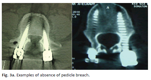
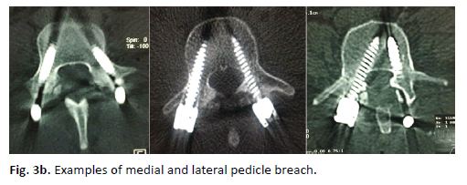
Screw placement was considered correct when the screw was completely surrounded by bone with no portion of the screw perforated outside the pedicular walls. Penetration of the wall of the pedicle was measured in millimeters using the scale on the CT image. Depending on the direction of the pedicle violation, the screw misplacement was noted as lateral, medial, inferior, or superior, and right or left.
Sample Characteristics
One hundred and three (103) patients were included in this study. Demographically, the study included 58 females forming a percentage of 56.3% and 45 males with a percentage of 43.7%. Mean and standard deviation for age were (45.92, 10.74) respectively, with a Confidence Interval (CI) of 95% (43.8, 48.02) for the entire sample.
Regarding screw insertion technique, the frequency and percentage distribution of patients in relation to the surgical technique used, showed that (40 patients, 38.8%) were operated using the percutaneous technique and (63 patients, 61.2%) were operated using the open technique. Concerning the number of screws specific to patients under question, the frequency and percentage distribution for the number of screws was (67 patients, 65.0%) had four screws, (34 patients, 33.0%) had six screws and (2 patients, 2.0%) had eight screws inserted. Using a weighed average (WA), the mean value for the number of screws and standard deviation were (4.74, 1.048) respectively.
Verification of Hypothesis 1
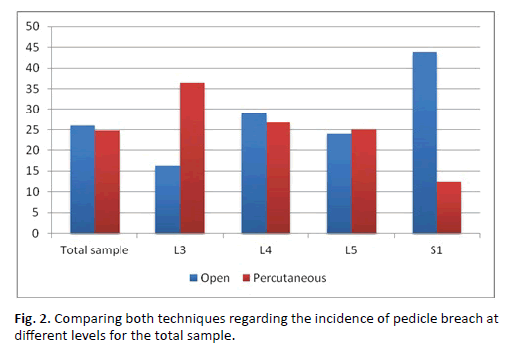
Testing the differences between the two techniques for the total sample and different levels: The following tables project the statistical results through which both techniques have been compared for the total sample and different levels using testing for proportions for two independent samples (Z) test (Table 1).
| Open technique (Op) | Percutaneous technique (Pr) | ||
|---|---|---|---|
| Pedicle n1 | % | Pedicle n2 | % |
| 71 | 26.0 | 45 | 24.7 |
Z=0.31 P-Val.=0.758 (P>0.05)
Table 1a: Incidence of pedicle breach according to technique used (Total sample).
| Open technique (Op) | Percutaneous technique (Pr) | Z | P | Comment | |||
|---|---|---|---|---|---|---|---|
| Pedicle n1 | % | Pedicle n2 | % | ||||
| L3 | 7 | 16.3 | 8 | 36.4 | 1.72 | 0.086 | Op=Pr |
| L4 | 32 | 29.1 | 16 | 26.7 | 0.34 | 0.735 | Op=Pr |
| L5 | 25 | 24.0 | 17 | 25.0 | 0.14 | 0.886 | Op=Pr |
| S1 | 7 | 43.8 | 4 | 12.5 | 2.28 | 0.023* | Op<Pr |
*Denotes (Z test) is sig. at 0.05 level of significance.
Table 1b: Incidence of pedicle breach according to technique used (Distribution according to levels).
Generally no statistical difference was found between both techniques regarding the presence of pedicle breach (main complication). This applied for all levels operated except the S1 level where a statistically significant difference was found in favor of the percutaneous technique at a 0.05 level of significance. Figure. 2 illustrates the results above.
Comparing both techniques regarding the side and direction of pedicle breach
Tables 2 and 3 project the statistical results through which both techniques have been compared regarding side and direction for the total sample using Pearson’s chi square test (testing for independency).
| Technique Side |
Open technique | Percutaneous technique |
|---|---|---|
| Right % |
40 Pedicles 56.3 |
34 Pedicles 75.6 |
| Left % |
31 Pedicles 43.7 |
11 Pedicles 24.4 |
Pearson Chi-Square (χ2) =4.404 df=1 Sig.= 0.036 (P<0.05)
Table 2: Comparison of both techniques regarding the side of pedicle breach.
| Technique Direction |
Open technique | Percutaneous technique |
|---|---|---|
| Medial % |
35 Pedicles 49.3 |
18 Pedicles 40.0 |
| Lateral % |
36 Pedicles 50.7 |
27 Pedicles 60.0 |
Pearson Chi-Square (χ2) =0.959 df=1 Sig.= 0.327 (P>0.05).
Table 3: Comparison of both techniques regarding the direction of pedicle breach.
Regarding the side of the screw breach, the results of Table 2 show that there is a statistically significant difference, regarding the percentage distribution of the side of pedicle breach (right or left), between both techniques, using Pearson’s chi square (P<0.05). In the percutaneous technique, the left side showed a lower incidence of pedicle breach compared to the right side, whereas in the open technique, the incidence of pedicle breach was nearly balanced between both sides.
Regarding the direction of the screw breach, the results of Table 3 show that there is no statistically significant difference, regarding the percentage distribution of the direction of pedicle breach (medial or lateral), between both techniques, using Pearson’s chi square (P>0.05). The results indicate a nearly balanced distribution between both techniques with reference to the direction of pedicle breach.
Verification of Hypothesis 2
Comparing the degree of deviation between both techniques
This hypothesis aims at studying the reasons resulting in the degree of deviation through a Two-Way ANOVA model, taking into consideration the Technique as the main factor and another group of variables including (level, side and direction) as the other factors.
The Two-Way ANOVA was used to explain the degree of deviation (deviation in millimeters), which is the yield of a set of factors (independent variables). The Two-Way ANOVA model with interactions was employed in three different models as follows:
Model 1
This model attempts to explain the differences of the deviation in millimeters (Dependent variable) through the technique, level and the interaction between both (Independent variables), taking into consideration, that only levels L3,4,5 & S1 were studied (Table 4), due to the small number of cases in levels L1 & L2 representing 3.4%, which is less than 4% of the total number of cases showing screw deviation.
| Source of variations | Sum of squares | df | Mean square | F-ratio | Sig |
|---|---|---|---|---|---|
| a. Techniques | 11.380 | 1 | 11.380 | -- | -- |
| b. Level | 0.344 | 3 | 0.115 | -- | |
| c. Interactions (a´b) |
7.000 | 3 | 2.333 | -- | -- |
| Model | 24.643 | 7 | 3.520 | 1.405 | 0.211 |
| Residual | 270.538 | 108 | 2.505 | (P>0.05) | |
| Total | 295.181 | 115 | -- | -- | -- |
Table 4: Two-Way ANOVA for deviation in millimeters against technique and level.
Table 4 shows the results of the Two-Way ANOVA concerning the study of deviation in millimeters through the technique and level. The table insures, that the model was not significant, where the F-ratio of the model is (1.405) with degrees of freedom (7,108), (P>0.05). Consequently, there is no significant difference between the deviation in millimeters for all components of the model (techniques, different levels and also the interaction between them) (Figure 3).
Model 2
This model attempts to explain the differences of the deviation in millimeters (Dependent variable) through the technique, side and the interaction between both (Independent variables), where the sides included right and left.
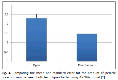
Figure. 4 and Table 5 shows the results of the Two-Way ANOVA concerning the study of deviation in millimeters through the technique and side.
| Source of variations | Sum of squares | df | Mean square | F-ratio | Sig |
|---|---|---|---|---|---|
| a. Techniques | 9.347 | 1 | 9.347 | 3.816 | 0.05 ** |
| b. Side | 4.431 | 1 | 4.431 | 1.809 | 0.181 |
| c. Interactions (a´b) |
0.380 | 1 | 0.380 | 0.155 | 0.694 |
| Model | 20.817 | 3 | 6.939 | 2.833 | 0.042** |
| Residual | 274.365 | 112 | 2.450 | ||
| Total | 295.182 | 115 | -- | -- | -- |
**Denotes that the F-ratio is significant at a 0.05 level of significance. (P<0.05)
Table 5: Two-Way ANOVA for deviation in millimeters against technique and side.
Table 5 insures, that the model was significant, where the F-ratio of the model is (2.833) with degrees of freedom (3,112), (P<0.05). By studying the results of all three components of the model, it was found, that the technique was the only significant component, beings significant at a 0.05 level of significance.
Multiple ranges tests (Multiple comparisons tests)
The differences between the two techniques were studied through TUKEY test-Post Hock test as shown in Table 6.
| Techniques | Open | Percutaneous |
|---|---|---|
| Open | 2.287(1), 0.216(2) | 0.817(3)* |
| Percutaneous | 1.470(1), 0.125(2) | |
| (1) Denotes the mean value . (2) Denotes the standard error . (3) Denotes the difference between two means . |
||
*Denotes a significant difference between the two techniques.
Table 6:Results of the multiple comparisons tests for the technique component of model 2 (Two-Way ANOVA).
The results of the TUKEY test as seen in Table 6, confirm the presence of statistically significant differences between both techniques. The statistical description represented by the mean value and the standard error for each technique separately confirms that these differences are in favor of the percutaneous technique regarding the degree of deviation, with less deviation occurring in the percutaneous technique.
Model 3
This model attempts to explain the differences of the deviation in millimeters (Dependent variable) through the technique, direction of deviation and the interaction between both (Independent variables), where the direction of deviation includes medial deviation and lateral deviation.
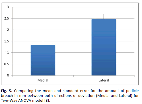
Figure. 5 and Table 7 shows the results of the Two-Way ANOVA concerning the study of deviation in millimeters through the technique and direction of deviation as well as the interaction between both.
| Source of variations | Sum of squares | Df | Mean square | F-ratio | Sig |
|---|---|---|---|---|---|
| a. Techniques | 18.248 | 1 | 18.248 | 8.828 | 0.004*** |
| b. Direction | 32.422 | 1 | 32.422 | 15.685 | 0.000*** |
| c. Interactions (a´b) |
5.552 | 1 | 5.552 | 2.686 | 0.104* |
| Model | 63.672 | 3 | 21.224 | 10.268 | 0.000*** |
| Residual | 231.510 | 112 | 2.067 | -- | |
| Total | 295.182 | 115 | -- | -- | -- |
***Denotes that the F- ratio is significant at a 0.01 level of significance (P<0.01).
*Denotes that the F- ratio is significant at a 0.1 level of significance (P<0.1).
Table 7: Two-Way ANOVA for deviation in millimeters against technique and side:
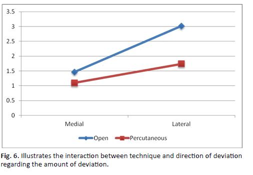
Table 7 insures, that the model was significant, where the F-ratio of the model is (10.268) with degrees of freedom (3,112), (P<0.01). By studying the results of all three components of the model, it was found, that the technique and direction were the significant components, at a 0.01 level of significance. Also, the results have confirmed, that the component specific to interaction came significant at a 0.1 level of significance only. Accordingly, the next step will be a detailed study of all three components of the model as follows:
Multiple ranges tests (Multiple comparisons tests)
The technique component of the model was not examined due to its previous examination in the previous model (being the main factor of the study). The differences between the two directions (Medial and Lateral) were studied through TUKEY test (Post Hock test) as shown in Table 8.
| Directions | Medial | Lateral |
|---|---|---|
| Medial | 1.341(1), 0.182(2) | 1.131(3)* |
| Lateral | 2.472(1), 0.207(2) | |
| (1) Denotes the mean value. (2) Denotes the standard error. (3) Denotes the difference between two means. |
||
Table 8: Predicted worst-case variable-level combinations for the different responses.
The results of the TUKEY test as seen in Table 8, confirm the presence of statistically significant differences between both directions. The statistical description represented by the mean value and the standard error for each direction separately confirms that these differences are in favor of the medial direction (the direction with less deviation) regarding the degree of deviation.
Studying the interaction component of the model:
The results of the Two-Way ANOVA Table 7 show, that the interaction component for the model (technique × direction) is statistically significant. Figure. 6 shows this interaction. In this figure, it can be seen that firstly there is a wide range of variation for the degree of deviation within the same technique. There are also obvious differences between both techniques regarding the amount of deviation concerning the lateral deviation (with lateral deviation being more in the open technique) in favor of the percutaneous technique. Lastly, there is a considerable degree of similarity between both techniques regarding amount of medial deviation, to an extent almost reaching intersection, between both techniques.
Statistical Conclusion
Studying the presence of pedicle wall violation in general, there is no statistical difference between both techniques. However, at the S1 level, there is a statistical difference in favor of the percutaneous technique. Regarding the side of violation, there is a lower incidence of pedicle breach on the left side in favor of the percutaneous technique. Regarding the direction of pedicle breach, there is no statistically significant difference between both techniques.
Studying the extent of pedicle breach in general, we found no effect of technique or level on the extent of pedicle breach. However, percutaneous technique had a lower amount of pedicle breach taking the side into consideration. The amount of medial deviation is smaller with the percutaneous technique.
Discussion
Pedicle screw fixation is superior to anterior fixation and posterior hook-rod fixation because the pedicle offers a strong point of attachment [1]. That’s why pedicle screw is widely used as a means of stabilization of the lumbar spine [11]. Wide range of indications for the use of pedicle screw exists, including trauma, deformity, tumors, infections, degenerative conditions and reconstruction. Recently, new techniques of screw insertion have made enormous progress, including the percutaneous technique of screw insertion. Proper pedicle screw insertion not only protects vital surrounding structures from injury but also it is important for long term survival of the metallic construct, in the term of better fusion and stronger construct [12].
The vital anatomic structures around the pedicle include the dural sac medially, the nerve roots superiorly and inferiorly, and the vascular structures anterolaterally. The risk of injury of these vital structures, according to Castro et al, 1996, occurs when the surgeon does not see the pedicle [3]. So, the safety and accuracy of pedicle screw fixation is crucial for the safety, efficiency and stability of the procedure [4]. For accuracy of pedicle screw insertion, it may be guided by anatomic landmarks and imaging, both preoperatively and intraoperatively. Imaging tools include plain radiography, fluoroscopy, and, more recently, image-guided technology [1].
Safety concerns and potential complications if screws are misplaced, causing pedicle wall disruption, have focused attention on screw placement techniques [5]. Since its introduction, accuracy of pedicle screws insertion has been the subject of many studies. A wide range of malpositioned screw rates have been reported [12]. The rate still may be considerable and has been reported to range up to nearly 40% [4]. Others reported the presence of unobserved malpositioned screw. In a review of the literature, a rate of 28.1% to 39.9% malpositioned pedicle screw in clinical studies and a 5.5% to 31.3% in cadaver studies were noted [13]. The percentage of malpositioned screws may be higher when normal anatomic landmarks have been obscured, as with revision surgery [14].
Fluoroscopic guidance intraoperatively demonstrates the depth of penetration not the malpositioned screw [15]. In cadaveric studies, direct observation of pedicle wall violation at dissection was the gold standard [7]. Asymptomatic violations of the walls of the pedicle still exist and can result in a weaker construct [6]. It is generally believed that CT imaging is superior to conventional x-rays in determining pedicle screw position, especially in the presence of pedicle wall violation. Moreover, it is superior regarding determination of the site of violation per pedicle [8]. However, no clear data exist on the sensitivity or the specificity of using CT images in identification of pedicle screw violation of the wall of the pedicle [16].
In practice, intra operative and postoperative assessment of pedicle screw fixation is mainly dependant on plain radiographs. In the present study we aimed at evaluating the incidence and accuracy of lumbar pedicle screw position, comparing the conventional and percutaneous techniques of screw insertion, using postoperative C.T guided evaluation, with 2 mm axial slices with bone window images, and in clinical terms evaluate whether minor displacements were responsible for clinical symptoms. Implant position and relation between clinical symptoms and radiological violation is reported. C.T assessment is usually reserved to cases of clinical doubt of pedicle wall violation or when symptoms of new neurological deficits appear [16]. However, for the sake of the study, C.T evaluation was done to detect even minor wall violations.
Review of literature considering the incidence of lumbar pedicle screw misplacement, revealed variable rates from 28.1% to 45% in clinical studies, with lower rates in cadaveric studies from 5.3% to 31.3% [14]. In reviewing results in the present study, according to technique used, the incidence of pedicle breach was 71 pedicles (26%) in open technique and 45 pedicles (24.7%) in percutaneous technique. This incidence of misplaced screws in our study is less than most published literature results.
Pedicle screw should be completely within the pedicle with no violation. Previous studies compared C.T. scans and plain x-rays of lumbar pedicle screw accuracy and documented 10 times definite violation than did plain x-ray [8]. In the present study, C.T. scan could detect minor pedicle wall violations even less than 2mm.
Fourth and fifth lumbar levels were the place of the highest pedicle wall violation in both techniques, in our study compared to L3.
This complies with other studies in the literature and is attributed to increased risk when more pedicular inclinations are present [5].
Generally, for all levels, except S1 level, this study concluded no statistical difference was found between open and percutaneous techniques regarding the presence of pedicle breach (main complication). For S1 level, there was a statistically significant difference in favor of the percutaneous technique at a 0.05 level of significance.
Powers et. al., 2006, reported that the incidence of screw displacement was lower in percutaneous technique than in open technique, and attributed that to the use of intraoperative fluoroscopy [10]. In the present study, we found no effect of technique or level on the extent of pedicle breach. However, percutaneous technique had a lower amount of pedicle breach taking the side into consideration. The incidence of lateral wall penetration in our study was slightly more common than medial violation in percutaneous technique while it was nearly the same in open technique. This complies with many results in the literature [3,4,12,16-18]. The amount of medial deviation is thus smaller with the percutaneous technique.
Regarding the side of the screw breach, there was a statistically significant difference, regarding the percentage distribution of the side of pedicle breach (right or left), between both techniques in the present study. In the percutaneous technique, the left side showed a lower incidence of pedicle breach compared to the right side, whereas in the open technique, the incidence of pedicle breach was nearly balanced between both sides.
Regarding the direction of the screw breach, medial and inferior violations are more likely to cause neurological deficits than superior and lateral violations being more toward the dura and existing nerve root [1,8,19]. In the present study, there was no statistically significant difference, regarding the percentage distribution of the direction of pedicle breach (medial or lateral), between both techniques. The results indicate a nearly balanced distribution between both techniques with reference to the direction of pedicle breach.
We attribute the reported difference between both techniques in favor of the percutaneous technique to the fact that respecting fluoroscopic anatomy of pedicle boundaries, and inserting the screw under strict fluoroscopic guidance in both medio-lateral and rostral-caudal orientations. Despite that, pedicle violations still do exists. With growing experience the incidence of pedicle screw violation definitely decreases.
Lastly, we concluded, together with other authors, that C.T. evidence of the presence and degree of pedicle wall violation, in conjunction with patient symptoms are probably the most impacted factors in determining the proper decision of further patient management protocols [1].
Conclusion
Pedicle screw insertion carries risk of pedicular wall violation even in experienced hands even with the use of intraoperative fluoroscopic guidance is used. However; most violations are minimal with no clinical consequences and can be evaluated best by C.T scan not plain x-rays. There is no statistical difference between open and percutaneous techniques, except at the S1 level, in favor of the percutaneous technique. Moreover, percutaneous technique had a lower amount of pedicle breach taking the side into consideration. The amount of medial deviation is smaller with the percutaneous technique.
REFERENCES
- Akyildiz F., Soyuncu Y., Yildirim F. B., et al.: Anatomic evaluation and relationship between the lumbar pedicle and adjacent neural structures an anatomic study. Spinal Disord Tech. 2005; 18: 243-246.
- Attar A., Ugur H. C., Uz A., Tekdemir I., et al.: Lumbar pedicle: Surgical anatomic evaluation and relationships. Eur Spine J. 2001; 10: 10-15.
- Castro WH., Halm H., Jerosch J., et al..: Blasius S.: Accuracy of pedicle screw placement in lumbar vertebrae. Spine. 1996; 21: 1320-1324.
- Gonzalez-Cruz J., Karim A., Mukherjee D., et al.: Accuracy of pedicle screw placement for lumbar fusion using anatomic landmarks versus open laminectomy: A comparison of two surgical techniques in cadaveric specimens: Operative Neurosurgery. 2006; 1: 13-19.
- Robertson PA., Stewart NR.: The radiologic anatomy of the lumbar and lumbosacral pedicles. Spine. 2000; 25: 709-715.
- Wang M. Y., Kim K. A., Liu C. Y., et al..: Reliability of three-dimensional fluoroscopy for detecting pedicle screw violations in the thoracic and lumbar spine. Neurosurgery. 2004; 54: 1138-1143.
- Grauer J. N., Vaccaro A. R., Brusovanik G., et al.: Evaluation of a novel pedicle probe for the placement of thoracic and lumbosacral pedicle screws. J Spinal Disord Tech. 2004; 17: 492-497.
- Ahlgren B. A., Learch T. J., Massie J. B., et al.: Assessment of pedicle screw placement utilizing conventional radiography and computed tomography: A proposed systematic approach to improve accuracy of interpretation. Spine. 2004; 29: 767-773.
- Haid RW., Morone MA.: spondylolisthesis and spondylolysis. In: George T. Tindall, Paul R. Cooper, Daniel L. Barrow (eds): The practice of neurosurgery. Williams and Wilkins. Baltimore, Meryland. 1997; 6: ch167, 2541-2563.
- Powers C. J., Podichetty V. K., Isaacs R. E.: placement of percutaneous pedicle screws without imaging guidance. Neurosurg Focus. 2006; 20: E3.
- Badran M., El-Ghandour N. M. F., Mohey M.: Comparative study between narrow dynamic compression plate with transpedicular screws and Titanium transpedicular fixation system in the surgical treatment of symptomatic nontraumaticspondylolithesis in adults. EJNS. 1999; 14-1: 175-180.
- Albert T. J., Grauer J. N., Vaccaro A. R., et al.: Evaluation of a novel pedicle probe for the placement of thoracic and lumbosacral Pedicle screws. Spinal Disord Tech. 2004; 17: 492-497.
- Glossop N. D., Hu R. W., Randle J. A.: Computer-aided pedicle screw placement using frameless stereotaxis. Spine. 1996; 21: 2026-2034.
- Austin M. S., Vaccaro A. R., Brislin B., et al.: Image-guided spine surgery;a cadaver study comparing conventional open laminoforaminotomy and two image-guided techniques for pedicle screw placement in posterolateral fusion and nonfusion models. Spine. 2002; 27: 2503-2508.
- Açikbas S. C., Tuncer M. R.: New method for intraoperative determination of proper screw insertion or screw malposition. J Neurosurg (Spine 1). 2000; 93: 40-44.
- Yoo J. U., Ghanayem A., Petersilge C., et al..: Accuracy of using computed tomography to identify pedicle screw placement in cadaveric human lumbar spine. Spine. 1997; 22: 2668-2671 .
- Li B., Jiang B., Fu Z., et al..: Accurate determination of isthmus of lumbar pedicle: A morphometric study using reformatted computed tomographic images. Spine. 2004; 29: 2438-2444.
- Tae K. L., Sang G. L., Chan W. P., et al.: Comparative analysis of adjacent levels of degeneration and clinical outcomes between conventional pedicle screws and percutaneous pedicle screws in treatment of degenerative disease at L3-5; A preliminary report. Korean J Spine. 2012; 9: 66-73.
- MohiEldin M., Arafa A. M.: Lumbar transpedicular implant failure: A clinical and surgical challenge and Its radiological assessment. Asian Spine J. 2014; 8-3: 281-297.




 Journal of Orthopaedics Trauma Surgery and Related Research a publication of Polish Society, is a peer-reviewed online journal with quaterly print on demand compilation of issues published.