Complications in treatment of older children with congenital clubfoot by Ilizarov external fixator
2 Scientific Institute for Traumatology and Orthopedics, Novosibirsk, Russia
Received: 26-Sep-2017 Accepted Date: Oct 06, 2017 ; Published: 09-Oct-2017
This open-access article is distributed under the terms of the Creative Commons Attribution Non-Commercial License (CC BY-NC) (http://creativecommons.org/licenses/by-nc/4.0/), which permits reuse, distribution and reproduction of the article, provided that the original work is properly cited and the reuse is restricted to noncommercial purposes. For commercial reuse, contact reprints@pulsus.com
Abstract
Background: The purpose of the study was to analyze and describe the complications that arose during the treatment of children aged between 7 and 17 years with congenital clubfoot in the Ilizarov Clinic using the transosseous osteosynthesis method as well as measures for their stopping and prevention.
Methods: In the Ilizarov clinic between 2005 and 2015, 108 patients (126 feet) aged between 7 to 17 years (mean age was 12.2 ± 2.3) with congenital clubfoot were treated. This group of patients was treated by the Ilizarov method with an original external fixator. In patients of this group along with insertion of wires in tibia and foot with the appropriate installation of Ilizarov frame, we used surgical interventions on soft tissues, osteotomies (V-shaped) and stabilizing surgeries (triple arthrodesis).
Results: In the course of treatment complications (inflammation, soft tissue cutting by a wire, premature consolidation of the osteotomy, subluxation in the ankle joint, cutting of the calcaneus by a wire) were observed in 22 patients, which amounted to 20.4% of the total number of patients. All encountered complications were typical for transosseous osteosynthesis and external fixators and were eliminated during treatment and did not affect its final result. In the long-term follow-up period (average 45 months), 93.5% of patients kept the desired result.
Conclusion: Abidance by the methodological principles of transosseous osteosynthesis by Ilizarov in the operative treatment of school-age children with congenital recurrent clubfoot and rational management of patients in the postoperative period is the prevention of complications and contributes to the achievement of the desired result of treatment.
Keywords
Clubfoot, External fixator, Ilizarov, Complications, Recurrence, Children Level of Evidence IV, retrospective study.
Introduction
Congenital clubfoot is one of the most common congenital malformations of the locomotor system which has a tendency to recurrence during child growth [1]. The prevalence of the disease is from 1 to 7 cases per thousand newborns [2], with 80% of these children growing in developing countries [3]. Older children have time to be treated repeatedly using a variety of techniques: serial casting, various surgical releases, using external fixation devices [4-14]. Some authors argue that the treatment of children with clubfoot by the Ponseti method is only relevant until the age of 9 [4]. Others describe the need for repeated more extensive intervention after primary surgical treatment. So, Lee notes the need for 35% of cases after surgery to produce a subsequent osteotomy of the foot bones [15]. Recurrences of clubfoot after various treatments according to the data of different authors range from 15% to 50% [5,7,8,11,14-16]. In children of 7 to years 17 years foot becomes more rigid after initial attempts of treatment of deformation. The treatment method by Ilizarov provides a stable controlled fixation of the foot bones, their gradual movement in the necessary direction and at a set pace stretching of shortened soft tissues, which allows the directional transformation of bone fragments and the foot as a whole [9,11,12,17-19].
Materials and Methods
The study is based on a retrospective analysis of the treatment process of 108 patients (126 feet) aged between 7 and 17 years (mean age was 12.2 ± 2.3) with congenital clubfoot between 2005 and 2015 at the Ilizarov Clinic. Minimum follow-up after surgery was 24 months (average 45 months, ranged from 24 months to 72 months). All patients previously received (most of them repeatedly) treatment in other hospitals. We used the clinical and X-ray examination (standing AP, ML, plantar AP views). Before treatment clinical evaluation of ankle joint and foot was done according to functional AOFAS (American Orthopaedic Foot and Ankle Society) scale [20], degree of deformation was determined according to the classification of A. Dimeglio [21]. In these children complex foot deformity was observed. Preoperatively the mean AOFAS was 32.7 ± 8.2 and the ankle motion was observed as 19° ± 11°. In these patients we used osteosynthesis method by Ilizarov (original frame). All patients received wires in the tibia and the foot with the appropriate assembly of the Ilizarov frame with hinge joints and an option of surgery on soft tissues and foot bones [9,12,17,18]. Surgical intervention on soft tissues included percutaneous z-shaped achillotomy, tenotomy of flexors of toes, capsulotomy of the metatarsophalangeal joints and plantotomy, transfer of m. tibialis anterior to 3rd cuneiform bone. We performed osteotomy of bones of mid foot and calcaneus (V-shaped), corticotomy of tibia and fibula for detorsion, and after 12 to years 13 years of age triple arthrodesis of the foot was produced. With gradual correction of foot deformities by the Ilizarov frame, existing scar-modified soft tissues were stretched, subluxations were eliminated in the joints of foot, all components of deformation of segment with hypercorrection of 10 degrees to 15 degrees were corrected.
Statistics was analyzed by means of the Attestat software (Attestation Software, Las Vegas, NV, USA). For the descriptive statistics, mean values of criteria and their standard deviation were defined.
The studies were approved by our institutional committee and were performed in accordance with the ethical standards laid down in the Declaration of Helsinki. All persons included into the study gave their informed consent and the study of human subjects followed the rules of clinical practice in the Russian Federation (RF Ministry of Health order # 266).
Results
Long-term results (average 45 months) of our treatment were analyzed by point-based system AOFAS. Among the long-term treatment results were distributed the following way: good in 72 patients (66.7%), satisfactory in 29 (26.8%), poor - 7 (6.5%). The mean AOFAS score increased significantly from 32.7 ± 8.2 preoperatively to 84.5 ± 6.1 postoperatively (p< 0.05). The ankle motion increased significantly from 19° ± 11° preoperatively to 29° ± 6° postoperatively (p< 0.05). In all cases, after conducted treatment improvement of the functional condition of the foot and its cosmetic view versus preoperative were noted. In the course of treatment complications (inflammation, soft tissue cutting by a wire, premature consolidation of the osteotomy, subluxation in ankle joint, cutting of the calcaneus by a wire) were observed in 22 patients, which amounted to 20.4% of the total number of patients. All the encountered complications were typical for transosseous osteosynthesis and were eliminated during treatment and did not affect its final result. Thus, 100% of patients in the treatment process achieved the desired result. However, it should be noted that in the long-term follow-up period (average 45 months) in 7 patients (6.5%) we had a recurrence of foot deformity due to incomplete compliance with recommendations and the tendency of the disease to recurrence, which required repeated intervention on the foot of these children in older age (triple arthrodesis of feet was performed).
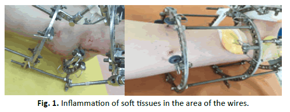
The most common complication was soft tissue inflammation around the wires which was noted in 8 patients (7.4%) and usually occurred when the aseptic and antiseptic rules were not followed. In most cases, this complication was observed on the inner surface of the feet, where the soft tissues experienced the greatest tension with distraction forces (Fig. 1). The clinical picture of inflammation was manifested by the corresponding general reaction of the body (increase in body temperature, leukocytosis, increased erythrocyte sedimentation reaction (ESR)) and characteristic local symptoms - pain, hyperemia, oedema of soft tissues in the area of entering or leaving the wires. To stop this complication infiltration of the inflammation zone by broad-spectrum antibiotics with novocaine, dressing with hypertonic solution and antibacterial ointments of local action (for example, chloramphenicol) were performed. In case of ineffective treatment within 2-3 days the wire was removed (2 cases). Prevention of soft tissue inflammation around the wires is compliance with the rules of asepsis and antiseptics, regular and high-quality dressing. In the first days of the postoperative period dressings were performed with the operating doctor every day, then weekly. Dressings were performed weekly by nurse. If it was necessary patient was observed in dressing room by surgeon.
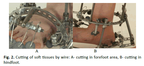
Another fairly common complication was the cutting of soft tissues by wires - 5 observations (4.6%). Cutting of soft tissues by wires was a typical wound process of compromised soft tissues of the foot inner surface in the area of the wires of the distal support which fixes the anterior or posterior part of the foot, with the subsequent possible attachment of infection (Fig. 2). In itself, the cutting of the skin by wires does not have a big effect on the treatment process, since the good regenerative capacity of the children’s skin ensures its rapid healing. The reasons for this complication were non-observance of the rules of carrying wires between the supports experiencing traction, non-observance of the tempo and the frequency of distraction, as well as the insufficiently stable fixation of the segment by the external fixator. To relieve this complication, the impact of distraction efforts in Ilizarov device was temporarily reduced or even stopped, the patient was recommended to temporarily restrain the weight-bearing on the operated extremity, we performed anti-ischemic and anti-inflammatory therapy. In order to prevent this complication, we recommend that the patient be given a course of restraining gymnastics of the foot and/or serial casting for 3-4 weeks, the wires should be carried with the satisfied stock of the skin between the supports which the traction forces will be directed to during the correction of deformation, and also the observance of the recommended rate and the frequency of the distraction effort required for a stable and rigid arrangement of the device.
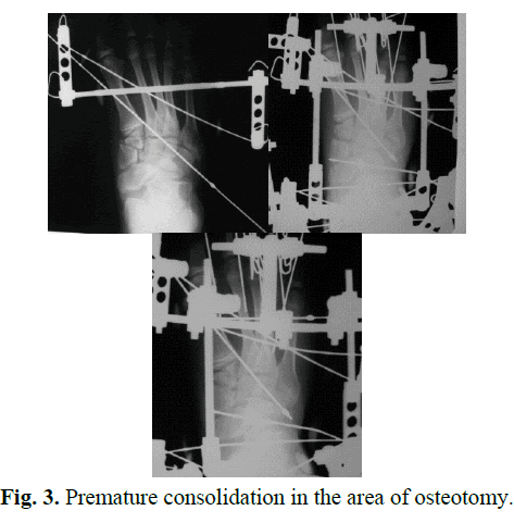
If the tempo and frequency of the applied compression-distraction forces were not followed and the age-related features of the foot osteogenesis were not taken into account, premature consolidation was received in the area of the performed osteotomy during correction of segment deformity (Fig. 3). This complication was clinically difficult enough to be suspected, which is connected with the possibility of the appearance of pain in the operated foot in varying degrees in response to the making compressiondistraction efforts and changes in the position of the foot. This is due to the individual threshold of the patient’s pain sensitivity. The complication was detected by X-ray and was noted in 5 patients (4.6%). To continue correcting the deformation of the foot in these cases, we needed to perform a weakening of the contact regenerate in the zone of consolidation. Accordingly, compliance with the tempo and frequency of distraction during treatment, timely and regular X-ray examination every 7 days to 10 days during the correction period will avoid premature consolidation in the area of osteotomy. In order to prevent this complication, we recommend that children of the young and preadolescent age begin correction after osteotomy from 4 days to 5 days after surgery, and patients of adolescence and older begin it from 6-7 days after surgery.
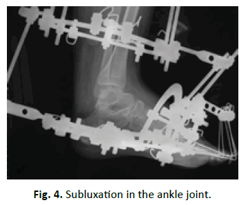
During correction of equinus component of foot deformity in 2 cases (1.9%) we identified a subluxation in the ankle joint anteriorly (Fig. 4), which was due to inaccuracies in the installation of hinges in the forefoot and inadequate control of the device functioning. Also in these cases, the wire was not carried through the talus. Clinically, oedema was observed on the anterior surface in the projection of the ankle joint and painful sensations in this area during weight-bearing. To eliminate this complication in the dressing room the Ilizarov device was changed with correct position of hinges and traction force to move the foot backward. The correction of subluxation was controlled by X-ray. The patient was recommended to limit the weight-bearing on the lower extremity within 2-3 days. Prevention of subluxation in ankle joint during correction of equinus component of foot deformity is the correct position of hinges, roads connecting the base in forefoot with the distal ring on tibia. When you perform osteotomy of foot in severe case (Dimeglio III-IV), it is necessary to fix the talus by olivial wire.
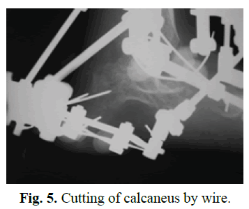
In two cases during correction of severe foot deformity cutting of bone by wire was noted, which developed as a result of the marginal pushing this wire through the calcaneus (Fig. 5). Clinically, this complication was manifested by the appearance of pain, oedema and hyperemia in the area of hindfoot and was confirmed by X-ray. Patients were recommended during this period a complete lack of weight-bearing on the operated foot. In both cases this complication arose at the end of the deformity correction period and required the removal of these wires and the repushing of new ones with appropriate installation of the support in the hindfoot. Prevention of the wire cutting of bone is the pushing of wires through the body of the calcaneus in different planes.
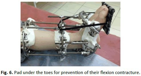
Special attention should be paid to trophic of forefoot soft tissues and foot sections experiencing the greatest tension. So, if the allowable value of a one-step intraoperative or postoperative correction is exceeded, it is possible to develop ischemic manifestations: blushing of the skin, local hypothermia, impaired sensitivity in the form of paresthesia, hypoesthesia or anesthesia, ischemic pain or even necrosis of the foot soft tissues. Thus, 5 patients after a one-stage intraoperative correction, due to ischemia of the forefoot, the traction efforts were eliminated, the position of the foot was returned to the original one and the deformation correction was performed gradually. To prevent flexion contracture and subluxation / dislocation in the joints of the toes, all toes were fixed by wires at the time of correction, after that they were fixed by pad under forefoot (Fig. 6).
All encountered complications were eliminated during treatment process and didn’t affect the final result of it.
After removal of the Ilizarov frame patient’s limbs were immobilized with a plaster cast from the upper part of the tibia up to foot toes in a position of hypercorrection of the foot up to 10-15° in all deformity components for the period of 1.5 months to 2 months. Patient was allowed to walk with gradually increasing weight-bearing on this extremity with or without crutches. After removal of the plaster cast all the patients were recommended to get restorative treatment in the domiciliary clinic including wearing of an orthopedic shoes with an insole-pronator, limb immobilization with an ankle-foot orthosis (AFO) for 6 months to 12 months for the night, massage and redressing exercises for the foot and also physiotherapy aimed to improve trophic of the anterior-external group muscles of the tibia, foot and restoration of the range of motions in the ankle joint. Amplitude of motions in the ankle joint and joints of toes was restored up to initial one on the average in 4 weeks to 6 weeks after the beginning of the active exercises.
Discussion
Nowadays, on the use of external fixators for correction limb deformity (and / or lengthening), the question is what is considered a complication and how to classify it [22-28]. There are a sufficient number of classification of complications [22,24-27], which are often encountered during treatment with transosseous osteosynthesis and they are in fact typical for this method. Accordingly, we can take into account these classifications in process of corrections of deformation both long and short segments by external frame. Paley suggests separating problems, obstacles, and sequela [24]. Cherkashin and colleagues define ‘complication’ as an unpredicted undesirable deviation from the treatment plan, which without appropriate resolution will lead to a failure to achieve treatment goals or to the development of a new pathology [25]. Dahl describes complications as mild, moderate and severe [26]. Lascombes offers classification for all bone elongation systems based on the criteria of a triple contract: gain in correction, projected treatment duration, and maintenance of musculoskeletal system function [27].
In our opinion, all complications can potentially be serious, since without appropriate attention and adequate measures they will lead to a change or interruption of treatment that will cause deterioration of the patient’s condition. Therefore, we agree with colleagues [25] that it is not necessary to divide complications into mild or moderate, obstacles and problems. In this publication, we describe the complications that we encountered in the treatment of children 7 years to 17 years old with congenital clubfoot by the method of transosseous osteosynthesis by Ilizarov without separating them by any degree and significance.
Most authors who use an external fixator in foot surgery in patients with clubfoot, often report cases of soft tissue inflammation around the wires or pins, and the incidence of inflammation varies from 15% to 100% of patients [11,13,14,19,29]. Infections in pin pathway are virtually inevitable in most patients treated by external fixation, and they typically respond readily to local care and oral antibiotics, but, if a pin-tract infection is ignored or inadequately treated, excessive pain, sepsis, and loss of fixation may all result [25].
In larger series of patients, surgeons describe the wider range of complications, such as fracture of the tibia, premature consolidation of osteotomy, soft tissue cutting, subluxation in the foot joints, vascular disorders, stiffness of foot, contracture of the toes and ankle, residual deformation [9,11-14,19].
Anyway, most authors using the Ilizarov method talk about complications during the fixator wear period [24-27]. Some surgeons showed us that in some cases a period of use of external fixator reached 14 months that is not rational and not adequate. In our opinion, such usage of the Ilizarov device can arise because of the wrong tactics of intervention and can discredit this method. In our study, the maximum period of use of the external fixator was 5.5 months.
Our percentage of unsatisfactory results (recurrence of deformity) with a large series of patients and the complexity of the pathology is not large enough (6.5%), which also confirms the successes in the treatment of this group of patients by external fixators according to reports of many surgeons [9-11,14,19]. Although, in the papers of some colleagues who used external fixator other results were reported: in Ferreira et. al study [11] in terms of observation on average in 58 months after treatment by Ilizarov frame recurrence of the deformity was found in 19 feet (50%), in Ahmed paper unsatisfactory results were found in 27,8% cases [30]. Freedman describes a recurrence of deformity in 11 patients out of 21 (52.4%) [16]. These facts about the recurrence of deformation confirm the complexity of the pathology and its tendency to recurrence. In our study we did not find residual deformity of the foot.
We hope that the measures described by us will help surgeons avoid or reduce the number of these complications that are typical for treatment by external fixators.
The main limitation of this study is that it is a retrospective analysis of the treatment of a very complex group of patients with severe and complex deformities of the feet and their tendency to recurrence.
Conclusion
Thus, the abidance by the methodological principles of transosseous osteosynthesis by Ilizarov in the operative treatment of school-age children with congenital recurrent clubfoot and the rational management of patients in the postoperative period is the prevention of complications and it contributes to the achievement of the desired result of treatment.
Conflict of Interest
There are no conflicts of interest.
REFERENCES
- Herring J.A.: Disorders of the foot. In: Herring JA ed. Tachdjian’s pediatric orthopaedics: from the Texas Scottish Rite Hospital for children. Vol 2. 5th ed. Philadelphia: WB Saunders; 2014. 761-883.
- Ponseti I.: Clubfoot: Ponseti management. 2nd ed. Global Help Publication; 2005. 31 p.
- Penny J.N: The neglected clubfoot. Techn Orthop. 2005;20(2):153-166.
- Alves C., Escalda C., Fernandes P., et al.: Ponseti method: does age at the beginning of treatment make a difference? Clin Orthop Relat Res. 2009;467(5):1271-1277.
- Haft G.R., Walker C.G., Crawford H.A.: Early clubfoot recurrence after use of the Ponseti method in a New Zealand population. J Bone Joint Surg. 2007;89:487-493.
- Agarwal A., Kumar A., Shaharyar A., et al.: The Problems Encountered in a CTEV Clinic: Can Better Casting and Bracing Be Accomplished? Foot Ankle Spec. 2016;9(6):513-521.
- Hsu L.P., Dias L.S., Swaroop V.T.: Long-term retrospective study of patients with idiopathic clubfoot with posterior medial-lateral release. J Bone Joint Surg Am. 2013;95(5):271-278.
- Ettl V., Kirschner S., Krauspe R., et al.: Midterm results following revision surgery in clubfeet. Int Orthop. 2009; 33:515-520.
- Kirienko A., Villa A., Calhoun J.H.: Ilizarov technique for complex foot and ankle deformities. Marcel Dekker Inc., New-York, 2004; 459 p.
- Ganger R., Radler C., Handlbauer A., et al.: External fixation in clubfoot treatment - a review of the literature. J Pediatr Orthop B. 2012;21(1):52-58.
- Ferreira R.C., Costo M.T., Frizzo G.G., et al.: Correction of neglected clubfoot using the Ilizarov external fixator. Foot Ankle Int. 2006;27(4):266-273.
- Leonchuk S.S., Neretin A.S., Ivanov G.P.: Surgical Treatment of the Older Age Children with Congenital Recurrent Clubfoot Using Method of Transosseous Osteosynthesis by Ilizarov. J Bone Rep Recomm. 2017;3:1
- Eidelman M., Keren Y., Katzman A.: Correction of residual clubfoot deformities in older children using the Taylor spatial butt frame and midfoot Gigli saw osteotomy. J Pediatr Orthop. 2012;32(5):527-533.
- Fernandes R.M., Mendes M.D., Amorim R., et al.: Surgical treatment of neglected clubfoot using external fixator. Rev Bras Ortop. 2016;51(5):501-508.
- Lee D.K., Benard M., Grumbine N., et al.: Forefoot adductus correction in clubfoot deformity with cuboid-cuneiform osteotomy: a retrospective analysis. J Am Podiatr Med Assoc. 2007;97(2):126-133.
- Freedman J.A., Watts H., Otsuka N.Y.: The Ilizarov method for the treatment of resistant clubfoot: is it an effective solution? J Pediatr Orthop. 2006;26;432-437.
- Ilizarov G.A., Shevtsov V.I., Kuzmin N.V.: Method of treatment of the talipes equinocavus. Orthopedics traumatology and prosthetics. 1983;5:46-48.
- Ilizarov G.A.: Transosseous osteosynthesis. Springer-Verlag, Heidelberg. 1992.
- Refai M.A., Song S.H., Song H.R.: Does short-term application of an Ilizarov frame with transfixion pins correct relapsed clubfoot in children? Clin Orthop Relat Res. 2012;470(7):1992-1999.
- Kitaoka H.B., Alexander I.J., Adelaar R.S., et al.: Clinical rating systems for the ankle-hindfoot, midfoot, hallux and lesser toes. Foot Ankle Int. 1994;15(7):87-101.
- Diméglio A., Bensahel H., Souchet P., et al.: Classification of clubfoot. J Pediatr Orthop B 1995:4;129-136.
- Caton J., Dumont P., Bérard J., et al.: Etude des resultants à moyen terme d’une série de 33 allongements des members inférieurs selon la technique de H. Wagner. Rev Chir Orthop. 1985;71:44-48.
- Naudie D., Hamdy R.C., Fassier F., et al.: Complications of limb lengthening in children who have an underlying bone disorder. J Bone Joint Surg Am. 1998;80:18-24.
- Paley D.: Problems, obstacles, and complications of limb lengthening by the Ilizarov technique. Clin Orthop Relat Res. 1990;250:81-104.
- Cherkashin A.M., Samchukov M.L., Birch J.G., et al.: Evaluation of complications of treatment of severe Blount’s disease by circular external fixation using a novel classification scheme. J Pediatr Orthop B 2015. 24:123-130.
- Dahl M.T., Gulli B., Berg T.: Complications of limb lengthening. A learning curve. Clin Orthop Relat Res. 1994; (301):10-18.
- Lascombes P., Popkov D., Huber H., et al.: Classification of complications after progressive long bone lengthening: proposal for a new classification. Orthop Traumatol Surg Res. 2012;98(6):629-637.
- Makhdoom A., Qureshi P.A., Jokhio M.F., et al.: Resistant clubfoot deformities managed by Ilizarov distraction histogenesis. Indian J Orthop. 2012;46(3):326-332.
- El-Mowafi H.: Assessment of percutaneous V osteotomy of the calcaneus with Ilizarov application for correction of complex foot deformities. Acta Orthop Belg. 2004;70(6):586-590.
- Ahmed AA.: The use of the Ilizarov method in management of relapsed club foot. Orthopedics. 2010; 33(12):881.



 Journal of Orthopaedics Trauma Surgery and Related Research a publication of Polish Society, is a peer-reviewed online journal with quaterly print on demand compilation of issues published.